The official account for the Life Science Solutions division of the Nikon Healthcare Business, with a focus on biological microscopy products and services.

Melville, NY
Joined April 2017
🧬Understanding #apoptosis is critical for studying cell health, drug responses, and disease progression.​ The ECLIPSE Ji enables you to streamline apoptosis assays using AI-driven imaging and analysis workflows. 📖 Read the application note: bit.ly/4zTfPgc
1
1
182
Introducing Detect ai in NIS-Elements v7 - Built on the Segment Anything Model, it turns hours of tedious manual annotation into simple clicks for both 2D and 3D images. Contact your Nikon rep to learn more👉 bit.ly/4Bnvc20 *This software version is available regionally
1
1
4
347
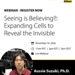
Join Nikon and Dojindo for a live webinar featuring Aussie Suzuki, Ph.D., Associate Professor at the University of Wisconsin–Madison. 📅November 10 🕑2:00 PM ET | 1:00 PM CT | 11:00 AM PT Register👉 bit.ly/4jwTsZd #ExpansionMicroscopy #CellBiology #Bioimaging
1
3
378
👋 See you at #Neuroscience2026 in Washington, D.C. Be sure to stop by Booth 1217 to meet the Nikon team. We look forward to connecting with you and discussing the latest advances in microscopy and imaging. #NikonMicroscopy #SfN #Neuroscience #Conference #Event @SfNtweets
239
🗣️"The ECLIPSE Ji-AX has transformed the way we work, combining intelligent automation with high-end imaging to make advanced microscopy accessible to everyone in the lab." Read the full interview with Professor Sylvain Gabriele: bit.ly/4eHH0mD
2
1
290
Our microscopy glossary covers 900+ terms, explained clearly and concisely, helping you quickly understand everything from basic concepts to advanced imaging techniques. 📚 Bookmark it here: bit.ly/4yt6jA8
247
A new study by Giulia Raffaele and colleagues at @IITalk, in collaboration with the Nikon Imaging Center #NIC, shows that rice roots change the way they grow in response to signals from the arbuscular mycorrhizal fungus Rhizophagus irregularis. 🔗 bit.ly/4dQAkBC
1
4
479
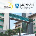
✨We are excited to announce that @MonashMalaysia Optical Imaging Platform (MUM OIP) has partnered with Nikon to establish the Nikon Center of Excellence #NCoE. Learn more about their available Nikon microscope systems👉 bit.ly/4yrMwBj
3
487
🔍 From rapid screening to high-resolution confocal imaging. In our interview, Professor Sylvain Gabriele of the University of Mons explains how the ECLIPSE Ji-AX helps his team investigate complex biological processes across multiple scales. Read more: bit.ly/4di44qR
3
387
🧪 With the fully automated Nikon ECLIPSE Ji, researchers can simplify cytotoxicity analysis using AI-driven imaging workflows that improve efficiency, consistency, and ease of use.​ 📖 Application note: bit.ly/4ACy8aD #NikonMicroscopy #ECLIPSEJi #CellAnalysis
2
378
📸 What a fantastic two days at the Nikon Microscopy Centers & Ambassadors Meeting Europe 2026! 💛A huge thank you to everyone who joined us and contributed to the inspiring discussions, presentations, and collaborations throughout the event.
7
505
We are excited to participate in #ASRM2026 from 24 - 26 October in Baltimore, Maryland! 👥 Stop by our booth 500 - the Nikon team will be happy to answer your questions ✅ See you there 👋 #NikonMicroscopy #ASRM
333
🔬🚀 Great to see NEMO in action aboard the #ISS! More on the Nikon technology behind this mission 👉 microscope.healthcare.nikon.… #LifeScience #SpaceResearch #NikonMicroscopy
We found NEMO! Here on the @Space_Station that stands for the newly installed Nikon Experimentation Microscope in Orbit (NEMO), designed and built by @NikonUSA and @BioServeST. This fresh addition to our scientific repertoire is not only a fully automated imaging system that allows for real time analysis, it also provides an incubator that controls environmental conditions (temperature, %CO2) to maintain live cells throughout the the process. This capability could lead to breakthroughs in drug discovery, tissue engineering and life science research. Photo 1: Image of the test sample from the first operation of the microscope. For you biology nerds out there, these are mesenchymal stem cells (GOLGI Bodies are stained green and microtubules are orange/red). Bio Art! Video: Installing the test sample prior to the imaging. Special thanks to @shankini at @BioServeST for the image (and amazing remote guiding for so many of our experiments on the ISS).
1
7
1,121
🧬A new Fluorescence Unmixing algorithm in NIS-Elements helps recover clean channel separation when using multiband filters for faster acquisitions without compromising image quality. Get in touch with us👉bit.ly/46H0LWv *This software version is available regionally.
1
4
411
The Nikon Microscopy Centers & Ambassadors Meeting Europe 2026 starts today! 🎉 Bringing together experts, innovators, and partners for two days of meaningful conversations and discoveries. Stay tuned for highlights. #NikonMicroscopy #LifeScience #Networking
2
354
In our latest application note, in collaboration with Julián Gurgo Bentivoglio, Erika De Sensi, and Alvaro Crevenna at EMBL Rome, a cyclic imaging workflow was used to detect and map 16 astrocyte-specific genes in mouse brain tissue. 📄Download: bit.ly/4dv4j1P
2
2
415
NIS-Elements Cluster Computing* offloads image analysis, AI training, deconvolution, and Smart Experiment assay processing to available computers across the network without needing additional NIS licenses. Contact us to learn more: bit.ly/4rpRGeb *Available regionally
1
4
404
We had a fantastic time at #ECP2026 - European Congress of Pathology! Thank you to everyone who visited our booth in Stockholm and joined us for great conversations throughout the congress. It was a pleasure showcasing our latest pathology and imaging solutions.
1
473
Nikon Microscope Solutions retweeted
Presenting... 🥁 ... The 2026 Nikon Small World in Motion winners! Congratulations to all of our winners, and thank you again to everyone who entered! The full gallery is available to view now: nikonsmallworld.com/gallerie…!
23
1
57
30,924