

Optica, Advancing Optics and Photonics Worldwide.
- Tweets20.5K
- Following1.5K
- Followers21.8K
- Likes6.5K




ALT A researcher working on the multiplexed detector system.

ALT Timon Schapeler (pictured) was part of the research team that developed the new approach, which extends photon counting to larger numbers of photons.

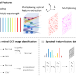
ALT Multiplexed optical feature extraction using spectral ensemble learning and spectral feature fusion. (a) Optical feature extraction setup: Light from a wavelength-tunable continuous-wave laser is directed onto a glass optical diffuser, generating distinct speckle patterns at each wavelength. (b) Spectral ensemble learning: classification of retinal OCT images. Speckle patterns obtained at different wavelengths are processed independently using linear regression. The predictions from each wavelength are then combined through majority voting to obtain the final classification across eight retinal disease categories. The consistency of predictions across different wavelengths also makes it possible to estimate uncertainty: strong disagreements indicate higher uncertainty. (c) Spectral feature fusion: data point separation. Speckle patterns obtained at multiple wavelengths are combined using weights that are adjusted during training to improve the separation of data points.

ALT A 3D cinematic rendering of the reconstructed elemental volume interacting with the structured X-ray beam. By applying a direct volumetric inverse problem to the data, copper (red), zirconium (green), and silver (blue) structures were accurately mapped with a 43-fold reduction in measurements compared with conventional raster scanning.

ALT A square image of C. elegans, a common model organism, seen under a microscope. Text reads International Day of Medical Physics, Advancing global health equity. The Optica logo is visible on the bottom left.


ALT A person speaking at a podium with a microphone, promoting the Optica Quantum 2.0 Conference and Exhibition, scheduled for 07–10 June 2027 at the Bella Center in Copenhagen, Denmark. The background is a muted color, and there's a call to "Submit Your Research." The Optica logo is displayed at the bottom.

ALT An image acquired using the new method (a) is compared with conventional radiology (b) under identical photon counts. [Image: Tiqiao Xiao, Shanghai Advanced Research Institute, Chinese Academy of Sciences]


ALT A square banner displaying various images of Paul Corkum. The text reads Congratulations 2027 Optica Honorary Member. Paul B. Corkum. University of Ottawa and National Research Council Canada.


ALT Two different 2-mm diamond chips with stamped GaP devices are glued to the cold finger of a cryostat to be cooled to 4 K. Sixteen sets of 40 nanophotonic cavities are arrayed over each chip.

ALT The optical ring-laser gyroscope uses a relatively large ring (pictured), which helped improve sensitivity.

ALT A person is holding a the flat gyroscope wafer with a grid pattern and a colorful edge. The grid contains various numbers marked on some sections.

ALT A purple electronic device labeled "Octave" on a perforated black surface, featuring various wires and components inside a rectangular frame.

ALT Two people stand in a lab next to equipment and a computer displaying graphs. One is dressed in a t-shirt, the other in a suit. The setting appears technical, involving scientific instruments.

ALT Helix Nebula

ALT The image shows the model’s simulations of color differences between observers viewing a Rec. 2020 capable display calibrated for the CIE 2006 2° standard observer. The upper left and lower right quadrants show the original color. The upper right quadrants show the color as seen by an observer with shifted sensitivity to red light. The lower left quadrants show the color as seen by a person with a shifted sensitivity to green light.