






Pinned Tweet
"BOW TIE". Papillary thyroid carcinoma. #FNA Thyroid. #Entpath #endopath #pathology #pathtwitter #PathTweetAward @MSWPathology @MyCytopathology @SamKhader

A Matloob, MD retweeted

“Vortex”, incipient tornado (or “onion skinning”), a subtle but helpful diagnostic feature of Papillary Thyroid Carcinoma. (FNA)

A Matloob, MD retweeted
“The Two Eyeballs”, located by my able fellow “John Sanchez” @JRS0298 in a case of Papillary Thyroid Carcinoma from today’s signout. (FNA)

A Matloob, MD retweeted
Textbook case, straight from the scope - “Squamous Cell Carcinoma of the Bladder” (Voided urine, ThinPrep)


A Matloob, MD retweeted
My “CASE of the week” for inhouse cyto signout, illustrating the remarkable phenomenon of how the “bag of polys” are recruited by the “endometrioid endometrial adenocarcinoma”, even at metastatic sites. This case is an abdominal fluid involved by the pt’s known EM CA.


A Matloob, MD retweeted
TWO, often overlooked cytomorphologic features of Medullary Thyroid Carcinoma on FNA - Intranuclear inclusions and focal nuclear pleomorphism/anisonucleosis.


A Matloob, MD retweeted
MYOEPITHELIAL cells stained with Giemsa on a parotid FNA are most photogenic. Although the DD here involves a “Myoepithelial Neoplasm”, other areas in the case had classic features of a “Pleomorphic Adenoma”.

A Matloob, MD retweeted

Pathology Residents: Biopsy of a cluster of umbilicated vulvar papules showing a classic example of? Extra credit for the name and intracellular location of the characteristic inclusions. #GYNpath #MedEd @nyulangone @nyugrossman @NYUGSOMpathres @NYUGSOM_Path


A Matloob, MD retweeted

DQ-stained cytospin preparation of pleural fluid with metastatic small cell ca. On the left you see a group of cells with ⬆️N:C ratio, finely granular chromatic, and molding. On the right you can appreciate the “single file”, “vertebral column”, or “stack of coins" arrangement.


A Matloob, MD retweeted

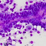
77 yo M w/ biliary stenosis. Bush #cytology during ERCP. No dysplasia, Giardia Lambdia 👻

A Matloob, MD retweeted

What are these gorgeous pink structures? Where are you most likely to find them (surprisingly, it's NOT sarcoidosis!)? Answer & more pics here: kikoxp.com/posts/5569. #pathology #pathologists #pathTwitter #dermpath #dermatology #dermatologia #dermtwitter

A Matloob, MD retweeted
Straight from the ultrasound room - Recurrent “ Fibrolamellar Carcinoma” (Fibrolamellar Hepatocellular Carcinoma). Liver FNA in a young boy.


A Matloob, MD retweeted
Pleural fluid in a patient with history of of #AML. It’s imperative to review the peripheral blood flow cytometry or smear in cases with abundant red blood cells. Some of these cells are suggestive of blasts; however, peripheral blood contamination cannot be excluded. #CytoPath


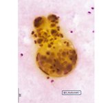
Lung tissue involved by Blastomycosis. GMS and PAS-F stains @SamKhader @SaeedAsiry @MSWPathology #FNA #blastomycosis #cyto #cytopath #path #pathology


A Matloob, MD retweeted
Interesting morphology of a squamous cell carcinoma metastatic to the pleural cavity (not common to see SqCC in an effusion #CytoPath specimen). Some of the cells have vacuolated and wispy cytoplasm. Note the combo stain for p40/DSG3. The lung tumor had a papillary architecture.


