

Annals of the Child Neurology Society is a peer-reviewed open access journal and an official #ANA_journals of @ChildNeuroSoc. Find us on LinkedIn too.
- Tweets176
- Following564
- Followers699
- Likes45





ALT Figure 2. Time to diagnosis and three clinical milestones in all participants.


ALT Figure 2. Graph of overall leukodystrophy patient volume (blue bars) and percentage of mortality (red line), mapped against individual hospitals (*deidentified) on the x-axis.

ALT Figure 1. Distribution of AMNL (adrenal insufficiency, myelopathy, neuropathy, leukodystrophy) scores at baseline visit in male patients of the Dutch adrenoleukodystrophy (ALD) cohort.


ALT Management Guidelines for the Treatment of Pediatric Brain Tumor–Associated Seizure

ALT Exploring Healthcare Continuity in Pediatric-Onset Multiple Sclerosis in the United States ACNS article screenshot

ALT "The Most Difficult Conversations We Have" ACNS article preview

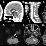
ALT Figure 1. Computed tomography of the brain and cervical spine at presentation demonstrated a calcified anterior craniocervical junction mass (A, arrow) with associated contrast enhancement (B,C, arrows). Magnetic resonance imaging demonstrated extensive cervical cord compression (D, arrow), no evidence of reduced or facilitated diffusion on apparent diffusion coefficient sequences (E, arrow), significant susceptibility weighted artifact (F, arrow), hypointensity on T2-weighted sequences without invasion of the adjacent jugular foramen (G, arrow), and avid enhancement on post-T1 weighted sequences (H, arrow).

ALT Dr. N. Paul Rosman (1934–2026)

ALT Reconciling the Use of Human Fetal Tissue in Research commentary in Annals of the Child Neurology Society

ALT Figure 4. Proposed model for cycle of brain processes contributing to prolonged stroke-like episode and SRSE in SWS. BBB, blood–brain barrier; SRSE, super-refractory status epilepticus; SWS, Sturge–Weber syndrome.


ALT Figure 1. Referral criteria for pediatric neuropalliative medicine clinic with categories of conditions seen in the first 5 years of the clinic and the types of medical decisions discussed during clinic visits.

ALT Dr. Peters in his home office in 2002.
