"Every image tells a story of love, hope, and new beginnings. #EchoTech"

Yemen
Joined June 2021
classic sign of ?
79
59
31
1,151
251,498
child presents with a prominent swelling over the left cheek. ultrasound reveal a well-defined, heterogeneous soft-tissue lesion, while Color Doppler demonstrates marked internal vascularity. What is the diagnosis based on these clinical and sonographic findings? #Ultrasound
2
3
23
1,026
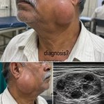
Patient 58-year-old man + slowly progressive, painless unilateral cheek swelling extending from the preauricular region to the mandibular angle + overlying skin remains smooth and intact, without erythema, ulceration, or a visible cutaneous punctum +
25
18
1
131
24,251
ultrasonography demonstrates a well-circumscribed, heterogeneous solid-cystic lesion containing multiple rounded hypoechoic compartments within the superficial soft tissues + Answer will be pinned after 48 hours.
1
4
3,477
the parotid-region location and characteristic multicystic internal architecture are the key clues to identifying the underlying salivary-region lesion. 🩺 Can you guess the diagnosis?
2
3
3,241
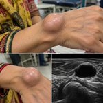
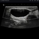
Patient 45-year-old + slowly enlarging, painless swelling over the dorsal wrist + well-circumscribed, smooth, dome-shaped, translucent subcutaneous mass with visible superficial vessels + no overlying erythema or ulceration
22
4
57
5,311
+ ultrasound demonstrates a well-defined, anechoic cystic lesion with posterior acoustic enhancement adjacent to the wrist joint, supporting a fluid-filled periarticular lesion + characteristic location and cystic sonographic appearance establish the key diagnostic clue.
2
1
2
925
🩺 Can you guess the diagnosis? Answer will be pinned after 24 hours.
1
672
Best ocular ultrasound pocket card 👌 #POCUS101 #ultrasound #POCUS @Pocus101
1
33
1
180
11,443
Patient 52-year-old presents with a slowly enlarging, painless, firm, nonmobile swelling in the supraclavicular fossa. The overlying skin is intact, without erythema or ulceration.
58
35
14
332
179,717
Ultrasonography reveals an enlarged, rounded, heterogeneous hypoechoic lymph node with loss of normal architecture. The absence of inflammatory changes and the suspicious nodal morphology warrant urgent evaluation for an underlying malignancy,
1
2
24
24,715
including thoracic and intra-abdominal primary tumors. The key diagnostic clue is a hard, painless supraclavicular node with suspicious ultrasonographic features. 🩺 Can you guess the diagnosis? Answer will be pinned after 24 hours. #Dermatology #MedEd #ClinicalDermatology
3
12
24,301
🩺 𝐍𝐔𝐓𝐂𝐑𝐀𝐂𝐊𝐄𝐑 𝐏𝐇𝐄𝐍𝐎𝐌𝐄𝐍𝐎𝐍 𝐕𝐒 𝐒𝐘𝐍𝐃𝐑𝐎𝐌𝐄 🩻 🔹 𝐖𝐡𝐚𝐭 𝐢𝐬 𝐭𝐡𝐞 𝐍𝐮𝐭𝐜𝐫𝐚𝐜𝐤𝐞𝐫 𝐩𝐡𝐞𝐧𝐨𝐦𝐞𝐧𝐨𝐧? The left renal vein is compressed, most commonly between the superior mesenteric artery (SMA) and the aorta.
1
6
40
1,853
♀️ 𝐏𝐞𝐥𝐯𝐢𝐜 𝐯𝐞𝐧𝐨𝐮𝐬 𝐜𝐨𝐧𝐠𝐞𝐬𝐭𝐢𝐨𝐧 in females 🔍 𝐊𝐞𝐲 𝐈𝐦𝐚𝐠𝐢𝐧𝐠 𝐂𝐥𝐮𝐞: Look for focal narrowing of the left renal vein at the point where it passes between the SMA and aorta, often with dilatation of the renal vein proximal to the compression.
1
1
145
💡 𝐊𝐞𝐲 𝐏𝐨𝐢𝐧𝐭: 𝐍𝐮𝐭𝐜𝐫𝐚𝐜𝐤𝐞𝐫 𝐩𝐡𝐞𝐧𝐨𝐦𝐞𝐧𝐨𝐧 = 𝐚𝐧𝐚𝐭𝐨𝐦𝐢𝐜𝐚𝐥 𝐜𝐨𝐦𝐩𝐫𝐞𝐬𝐬𝐢𝐨𝐧 𝐍𝐮𝐭𝐜𝐫𝐚𝐜𝐤𝐞𝐫 𝐬𝐲𝐧𝐝𝐫𝐨𝐦𝐞 = 𝐜𝐨𝐦𝐩𝐫𝐞𝐬𝐬𝐢𝐨𝐧 + 𝐜𝐥𝐢𝐧𝐢𝐜𝐚𝐥 𝐦𝐚𝐧𝐢𝐟𝐞𝐬𝐭𝐚𝐭𝐢𝐨𝐧𝐬 🩸 #radiology #radiologyInteresting #ultrasound #Ultrasound
1
2
134
Acute viral hepatitis in a young boy
2
3
45
2,346
4 months boy , came with Inguinal swelling , your opinion??
4
5
34
5,664
Acute pancreatitis
1
5
26
1,165