

Author. Consultant. Former #UCSB Director, Microscopy Facility. #UCLA alum. #microscopy - Photos & video are Copyright.
Southern California
Joined December 2016
- Tweets40
- Following338
- Followers281
- Likes18




Pinned Tweet
This silica shell was generated from a one-celled animal: a radiolaria. #microscopy #microscope #biology

Medusa obelia jellyfish1 mm diameter stained w/ fluorescent dye which specifically bind to protein #biology #microscopy #microscope #science

Introduction to Digital Photomicrography. Available thru Crowood Press and Amazon. #microscope #microscopy #science #photography #SciArt

Diatom photo with Reichert Anoptral contrast & processed with differential hysteresis algorithms. #microscope #microscopy #science #diatom

Montage showing human epithelial cells. What you record varies with the brand and type of microscope you use. #microscopy #biology #science

1
Triceratium morlandii diatom (from New Zealand) - a fossil species 32-40 million years old. #diatom #microscopy #fossil #science

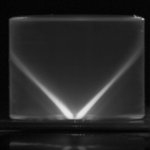
Light’s path to the specimen when using a darkfield condenser. #Thursdaytweet #microscope #microscopy #science #light #experiments

3
Diatom photo w/ Reichert Anoptral contrast & processed w/ differential hysteresis algorithms. #tuesdaytweet #microscope #microscopy #science

#Diatom illuminated with an antiquated lighting system - Austrian company Reichert's modified phase contrast system. #microscope #biology

A #photograph of an inverted #microscope—so named because the #optics for viewing the specimen are beneath the slide, not above it. #science


Klaus Kemp #diatoms slide. Modified my color Heine condenser & adjusted the light intensity for bicolor darkfield illumination. #microscope

Stained section of developing bone at the level of the epiphyseal plate. Used a #Tucsen DigiRetina 16 4K camera. #science #microscope #photo

#Photo of #diatom N. lyra. Its center looks like an outline of a lyre. Phase contrast microscope enhances contrast. #microscope #sciart

These are the shells of an algae - #diatoms. Their silica shell is perforated by pores arranged in geometric patterns. #biology #microscopic

The cellular constituents of these #diatoms were dissolved w/ strong acids leaving behind a silica shell. #microscope #microscopic #science

Camera info for this photograph on my Facebook page: facebook.com/optics4fun/phot… #camera #photograph #microscope #science

2
Circular arrangement of #diatoms taken with a Heine condenser. Info on #prismatic effect: facebook.com/optics4fun/phot… #microscope #science

