ECG Educator | Electrophysiologist | Professor @The_BMC | Alumni @BrighamFellows & @NYUMed | All tweets are my personal opinion 🩺

Boston, MA
Joined December 2019
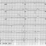
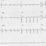
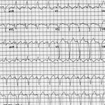
Patient with a history of palpitations. First ecg is baseline. Second ecg obtained 20 minutes later. #PodridECGs #ECG #meded #cardiotwitter
2
2
12
2,106
Patient with palpitations. What does the ECG show? #podridECGs #ECG #meded #cardiotwitter
4
3
9
1,915
Rhythm is regularly irregular with a sinus complex followed by an early qrs complex that has a negative p wave in leads 2 and aVF. These are PACs in a bigeminal pattern. At end is NSR.
1
1
91
Pt presents with episodes of lightheadedness. What does the ecg show? #PodridECGs #ECG #meded #cardiotwitter
5
2
4
894
Coarse AF (atrial waves are irreg in morphology, amplitude and intervals) with QRS complexes at a regular rate, ie regularized AF which is due to complete heart block. The escape rhythm is junctional and hence the CHB is due to AV nodal block.
2
118
Patient with known palpitations. Pulse is regular with occasional pauses. What does the ecg show? #PodridECG #ECG #meded #cardiotwitter
5
5
19
2,087
Atrial tachy with 5:4 Wenckebach. Nonspecific ST-T wave changes.
1
83
56 yo presents with palpitations. The ecg is obtained and what does it show? #PodridECGs #ECG #meded #cardiotwitter
5
1
2
657
Narrow complex regular Supravent tachy arrhythmia at a rate of 270 bpm. The only supravent rhythm at this rate is atrial flutter with 1:1 conduction. Also noted is electrical or QRS alternans. The j point depression and st depression may be due the superimposed flutter wave.
2
136
Patient with a history of palpitations. What is the etiology? #PodridECGs #ECG #meded #cardiotwitter
3
3
11
1,866
short rp supraventricular tachycardia. This is not sinus but may be junctional or atrial. Since it terminates with a non conducted p wave this indicates that this is a junctional tachycardia. Hence this is a typical avnrt.
2
111
74 yo man with an ischemic cardiomyopathy and a pacemaker complains of palpitations. What does the ecg show? #PodridECGs #ECG #meded #cardiotwitter
4
3
9
1,281
Biventricular pacemaker. It is tracking an atrial tachycardia that has a rate of 220 bpm. Pacemaker function is A sensed V paced but as atrial rate is greater than upper rate limit of pacemaker only every other p wave is sensed and hence vent pacing rate is 110 bpm.
1
3
156
76 yo man presents with episodic lightheadedness and presence. What does the ecg show? #PodridECGs #ECG #meded #cardiotwitter
4
2
14
1,861
At end of the ecg there is NSR with 2:1 block (which can be either mobitz type 1 or type 2. QRS has lbbb. At beginning there is chb with an escape ventricular rhythm. Hence the 2:1 av block is mobitz type 2.
1
144
34 year old woman with history of palpitations of undetermined etiology. This is her baseline ECG. What does it show and what is likely the etiology of the palpitations? #PodridECGs #ECG #meded #cardiotwitter
7
3
19
2,177
NSR with WPW. Note at end PR interval shortens and delta wave more prominent. This is termed “concertina effect” resulting from changes in av conduction velocity-when faster less preexcitation and when slower more preexcitation. Most frequent arrhythmia with WPW is avrt.
1
2
182
73 year old with know hypertension. No symptoms. What does the ECG show? #PodridECGs #ECG #meded #cardiotritter
7
5
24
3,667
The ecg provides evidence of myocardial abnormalities or hypertrophy and not chamber size or enlargement. This is diagnosed by an echo other imaging such as x-ray, ct scan or mri. Hence enlargement is not an appropriate ecg term.
1
70
NSR with narrow, tall and peaked p waves in leads 2,3 and aVF termed p pulmonary c/w right atrial hypertrophy or abnormality (not enlargement). Very neg p waves in leads v1-2 due to left atrial hypertrophy (not enlargement). Therefore biatrial hypertrophy or abnormality.
1
1
154