

🧠Neurosurgeon-Scientist | AI, Brain Cancer and Digital Health Researcher | Ambidextrous | Author |🇳🇵🇬🇧 @thecrick @kingsneuro @kingsimaging @naamii_nepal
London, UK
Joined March 2012
- Tweets404
- Following990
- Followers1K
- Likes6.1K





Pinned Tweet
“🚨 Podcast 🚨”
Dive deep into the world of neuro-oncology with ‘Neuro-oncology Talks | The Deep Dive.’ Explore groundbreaking research, AI in medicine, and the future of brain tumor treatment. 🎙️
Listen now: podcasts.apple.com/gb/podcas…
#NeuroOncology #CancerResearch #AIInMedicine

Prajwal Ghimire retweeted

As our brain ages, there is disruption of the flow of cerebrospinal fluid during sleep @NatureAging
nature.com/articles/s43587-0…

Prajwal Ghimire retweeted

Researchers have published the world's most detailed atlas of basal ganglia, comprising 25.5 million brain cells
go.nature.com/4C1XQpP

Prajwal Ghimire retweeted

Going from cold to hot.
How to get immunotherapy to work even better to treat cancer via the tumor microenvironment
@ScienceMagazine
science.org/doi/10.1126/scie…

Prajwal Ghimire retweeted

Even if you’re in it for the LONG haul, sometimes it’s good to come up SHORT!
If you can’t summarize, you’ll never memorize!
Here's a shortened, summary cheat sheet for my popular thread EVER—How to read a head CT
If you're looking at head CTs, this is what you NEED to know!
➡️3 main things to look for on head CTs are:
🔸Blood
🔸Mass Effect
🔸Stroke
➡️Blood
🔸CT is just a measure of density & blood is thicker than water, so blood is denser than brain on head CT
🔸Blood can be:
▪️EPIdural: looks lentiform
▪️SUBdural: looks crescentic
▪️SUBarachnoid: looks snake-like
➡️Mass effect
🔸You won’t bleed out in your head—the problem is the calvarium is an enclosed & brain gets compressed
🔸Look for:
▪️Subfalcine herniation = midline shift, ventricles to the other side
▪️Transtentorial herniation = loss of the basilar cisterns, normally look like a pentagon & smiley face
➡️Stroke
🔸Normal gray-white differentiation looks like an octopus & is crispy & well-defined
🔸In stroke, it looks like someone took a painting & smeared the white matter paint into the gray matter paint
🌟You can see the full thread here:
x.com/teachplaygrub/…
So now you know what to look for on a head CT. Hopefully this short version stays with you for the long haul!

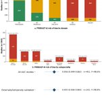
1/6 🧵 AI in neurosurgery is booming, but behind high statistical performance lies a critical clinical translation gap.
👉Here is what our largest meta-analysis of neurosurgical AI studies (n=1,063) published in @BJNSNeuro actually reveals 👇


5/6 📉 The Translation Gap
• 63.9% stopped at initial model development.
• Only 4.9% (52 studies) created a usable tool or prospectively evaluated models.
• 0 studies conducted an impact/implementation study.
• Just 0.3% reached regulatory clearance or routine deployment.

1
2
75
6/6 💡 The Takeaway
🩺To move from algorithm to bedside, AI research in neurosurgery must shift from publishing in-sample AUCs toward external validation, calibration reporting, and TRIPOD-AI/PROBAST-AI guidelines. @BJNSNeuro
📖 Open Access Paper: doi.org/10.1080/02688697.202…

3
71
Prajwal Ghimire retweeted
1/Do radiologists sound like they are speaking a different language when they talk about MRI?
T1 shortening what? T2 prolongation who?
Here’s a translation w/an introductory thread to MRI.

Prajwal Ghimire retweeted
1/Radiologist not answering the phone?
Just want a quick read on that stat head CT?
Here's a little help on how to do it yourself w/a thread on how to read a head CT!

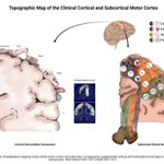
Our paper on clinical cortical and subcortical map of motor homunculus from @KingsNeuro has been included in 100 years in 💯 papers of @The_SBNS centenary celebration publication.
Thank you @The_SBNS.


4/6
2021-Ghimire P, Lavrador J, Baig A, Furlanetti L, Brogna C, Gullan R, Vergani F, Bhangoo R, Ashkan K. Intraop mapping of pre-central motor cortex and subcortex... Brain Struct Funct. 2021 Jun;226(5):1601-1611.
🔗doi.org/10.1007/s00429-021-0…
👏@prazNeuro

Prajwal Ghimire retweeted

Fantastic illustrations! Very useful to understand the concepts! @PrazNeuro
3/3 ⚠️ Surgical Safety:
👉Complications: in 48.9% , emphasizing the procedural challenge of deep-seated ventricular corridors.
👉Decisions should tailor the approach to specific tumour biology and patient presentation
Illustrations @PrazNeuro 👏


The choice of surgical technique alone does not govern complete resection or CSF diversion success. Instead, individual patient presentation, anatomical location, and tumour biology should guide the tailored selection of surgical approach. 👇
Excellent team effort @KingsNeuro 👏


Hot off Press:📜
1/3 🧵 How do conventional craniotomy and minimally invasive surgery (MIS) compare for intraventricular tumours?
A systematic review and individual patient data meta-analysis of 1,500 patients (361 studies) provides key clinical insights. 👇
@AsfandBm


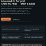
The Advanced 3D Neurosurgical Anatomy Atlas (brain & spine) is now public.
Open Access | Free
Any browser, 3D, VR and mobile version
10 interactive specimens, 34 guided surgical approaches, Rhoton Collection plates, white matter dissection walkthrough.
prazg.github.io/3D_Neurosurg…

Prajwal Ghimire retweeted

BREAKING NEWS
The 2026 #NobelPrize in Physiology or Medicine has been awarded to Karl Deisseroth, Peter Hegemann and Georg Nagel “for their discoveries concerning light-gated ion channels and optogenetics.”

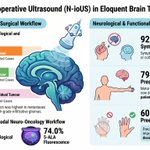
While navigated intra-op ultrasound offers a low-cost, real-time tool to compensate for dynamic intraoperative brain shift, its greatest standalone utility may be in lesions with purely anatomical endpoints, such as brain metastases.
@KingsNeuro @lavrador_jose
Infographic 👇

1/4 🧠 How do neurosurgeons maximize tumour cytoreduction while safeguarding neurological function in eloquent brain regions?
A study of 104 patients @KingsCollegeNHS evaluated navigated intraoperative ultrasound (N-ioUS) integrated into a modern multimodal workflow:👇
@feras199

Prajwal Ghimire retweeted

NEW Viewpoint: The clinician–artificial intelligence scientist: a proposed career pathway in medicine.
Read it here: thelancet.com/journals/landi…

Prajwal Ghimire retweeted
Waving the white flag when it comes to white matter anatomy?
Turns out white matter anatomy isn’t black & white!
Diffusion tensor imaging or DTI can help us to see the different systems in the white matter.
But often we don’t have DTI on routine brain MRIs.
So how can we tell if pathology is affecting important white matter functional anatomy?
We have to estimate, based on landmarks
Here’s how to remember the major tracts:
Motor tracts/Posterior Limb of the Internal Capsule/Corticospinal tract:
Corticospinal tract goes through your CORE. What is central in the brain? The ventricles!!
It runs along the lateral aspect of the lateral ventricles
Corpus Callosum:
Letter C looks like the curves of the splenium and genu of the corpus callosum
Superior Longitudinal Fasciculus:
What is superior & long in your body—your arms! Superior longitudinal fasciculus looks like arms hanging down along the lateral aspect of the brain
Inferior fronto-occipital fasciculus & Inferior longitudinal fasciculus
What’s inferior & long in your body—your legs! These curve around posteriorly and inferiorly, like two legs with their heels touching
So now you can always find the major white matter tracts!
Hopefully, now you can be right about what’s white!
