Pathologist, Dermatopathologist, UCLA Dept. of Pathology. Views are my own.
Joined February 2020
- Tweets15
- Following87
- Followers165
- Likes102
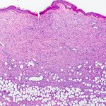

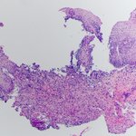



Dermatofibrosarcoma protuberans (DFSP) with a typical honeycomb appearance. This one shows entrapment of a few nerves (largest 0.3 mm), otherwise classic DFSP. Diffusely positive for CD34 (image not shown). #dermpath #PathTwitter #PathX #BSTpath


Keratin stain rarely disappoints and can save a life. When in doubt, just order. Here’s a Plasmacytoid Urothelial Carcinoma with no surface carcinoma in situ, supported by GATA3 after Keratin. Tumor cells remarkably mimic lymphoplasmacytic infiltrate. #PathTwitter #GUPath #PathX


Kaposi sarcoma in esophageal biopsy, immunocompromised patient. Looks like granulation tissue from low power.


Hx of oral erosions & flaccid body blisters. Classic example of Pemphigus Vulgaris: suprabasal acantholysis, tombstone sign: basal cells have lost lateral intercellular bridges, but remain attached to basement membrane. Intercellular IgG & C3 on direct IF. #dermpath #PathTwitter


If stuck between pigmented AK or early lentigo maligna on elderly head & neck, PRAME may be helpful. Especially if you tried a leaky Melan-A. See Santandrea et al. letter. I found it interesting and may try next time. #dermpath #PathTwitter
journals.lww.com/amjdermatop…

CMV in an immunosuppressed patient with esophageal ulcer. Pretty remarkable on the H&E and confirmed by IHC. CMV loves endothelial cells, you can see “megalo” cells in two capillaries at least. #PathTwitter #GIPath


Non-healing white patch on glans, older man. Differentiated penile intraepithelial neoplasia (PeIN). I find basal layer atypia most helpful. Note also dyskeratosis/ premature keratinization & ample eosinophilic cytoplasm; p53 null-type, slide not available. #dermpath #PathTwitter


Multiple perianal papules, rule out anal dysplasia. Turned out condyloma lata. Psoriasiform lichenoid reaction pattern with many plasma cells, clues to think of syphilis. Warthin-Starry worked very well highlighting spirochetes. IHC was positive. #dermpath #PathTwitter #syphilis


Here’s an example of how bland desmoplastic melanoma can look. It can be easily missed at scanning magnification. Here, residual melanoma is hiding under and blending with the scar. See nuclear crowding & haphazard growth in melanoma versus scar. #dermpath #PathTwitter #melanoma