

Cancer Systems Biology
Weizmann Institute of Science
Joined July 2019
- Tweets156
- Following136
- Followers2.3K
- Likes64




Tirosh Lab retweeted

IDH-mutant gliomas often progress slowly, but inevitably recur and progress. Are there spatial organizing principles at the level of cell states that repeat across tumours during stages of tumour progression?
cell.com/cancer-cell/fulltex…
Out this week in @Cancer_Cell, our work with @RouvenHoefflin, @TiroshLab, @MarioSuva, @GaliliNoam, and Christopher Mount quantitatively maps two independent axes of tumour organization, revealing how they emerge at different stages of progression and in distinct tissue contexts. We hope that understanding these recurring spatial patterns and associations will reveal new opportunities to halt tumour progression.

Tirosh Lab retweeted

🧵1/ Happy to share our new study:
"Spatial analysis reveals the evolving organization of IDH-mutant glioma"
We asked: How is spatial organization established in glioma before the hypoxic architecture of advanced disease emerges?
cell.com/cancer-cell/fulltex…

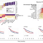
ALT Graphical Abstract
A collaborative effort between our lab at @WeizmannScience and the Puram Lab (@SidPuram) at @WUSTLmed, co-led by Dor Simkin and Tom Barrett. Many thanks to all authors for their contributions!
#SpatialTranscriptomics #HNSCC #Visium #CODEX #EMT (14/14)
Take-home model:
Tumor edge → fibroblasts / TGFβ → interface p-EMT
Tumor core → myeloid cells / OSM-OSMR → intratumoral p-EMT
Spatial context can separate biologically distinct versions of a state that may look similar in dissociated or bulk data. (13/14)
One important result was convergence: despite different locations, neighboring cells, and candidate signals, core and edge p-EMT showed highly similar expression programs.
Distinct microenvironmental interactions can therefore converge on a similar malignant phenotype. (12/14)
In two HNSCC cell lines, OSM induced EMT-associated and inflammatory transcriptional responses.
Together, the data support a model in which myeloid-derived OSM promotes p-EMT in tumor cores, alongside the established fibroblast-TGFβ mechanism at tumor edges. (11/14)
What could induce p-EMT in the core?
Ligand-receptor analysis pointed to OSM-OSMR:
• OSM was expressed mainly by immunosuppressive macrophages and neutrophils.
• OSMR was expressed by p-EMT malignant cells. (10/14)

CODEX provided single-cell, protein-level validation.
p-EMT core tumors contained p-EMT cells deeper within tumor nests and >10-fold more intratumoral pro-tumor myeloid cells.
p-EMT cells were closer to myeloid cells in core tumors, but to fibroblasts in edge tumors. (9/14)

The two patterns were embedded in different ecosystems:
• p-EMT edge: preferential association with fibroblasts at the boundary.
• p-EMT core: association with the Inf-Resp myeloid niche, increased intratumoral myeloid infiltration, and relative lymphocyte exclusion. (8/14)

Was this a sampling effect?
LAMC2 staining of 2-4 separate cores from 100 tumors showed that patterns tended to recur across regions of the same tumor.
p-EMT edge versus core therefore appears to be a tumor-wide spatial phenotype. (7/14)

The central surprise came from mapping p-EMT across tumor zones.
Some tumors showed the expected enrichment at the invasive tumor-stroma interface (“p-EMT edge”).
Others showed p-EMT deep within malignant nests (“p-EMT core”). (6/14)

The Inf-Resp program therefore captured more than abundance: it marked a spatial interaction between tumor-promoting myeloid cells and malignant cells.
Its gene signature was also associated with worse overall survival in an independent, combined cohort of 887 patients. (5/14)

We defined 16 recurrent spatial metaprograms representing individual malignant states and immune/stromal cell types.
One exception was an inflammatory-response (Inf-Resp) “micro-niche”: immunosuppressive macrophages and neutrophils closely associated with malignant cells. (4/14)
We mapped 26 HPV-positive and HPV-negative HNSCC tumors using 10x Visium, with matched scRNA-seq for 11 tumors.
We then tested key findings using Visium HD, LAMC2 IHC in 100 tumors, 31-plex CODEX in 40 tumors, and perturbations in 2 HNSCC cell lines. (3/14)

Our earlier work placed p-EMT at the leading edge of HNSCC, near CAFs, and showed that TGFβ can induce this program.
But is p-EMT always confined to the edge? Could the same state arise elsewhere through different microenvironmental interactions? (2/14)
cell.com/cell/fulltext/S0092…
Excited to share our new paper in @NatureGenet!
Using spatial transcriptomics and proteomics, we uncover two tumor-wide modes of partial EMT (p-EMT) in head and neck cancer, at the tumor edge or core, with distinct microenvironmental drivers. 🧵 (1/14)
nature.com/articles/s41588-0…

Registration for the Gordon Research Conference (GRC) in Single-cell cancer biology is now open! It will take place March 22-27, 2026, in Ventura, CA.
For more info and registration:
grc.org/single-cell-cancer-b…
Congrats Alissa!!
Highly recommended for students

I'm thrilled to share that I'll be starting my lab (greenwald-lab.org) at the Lunenfeld-Tanenbaum Research Institute @SinaiHealth and joining @MoGen_Grad at the University of Toronto as an assistant professor this fall. 1/
Tirosh Lab retweeted

👉The Curated Cancer Atlas.
Impressive: “124 scRNA seq datasets for >40 cancer types, with improved data annotations, visualizations and exploration”.
From Michael Tyler and @TiroshLab
@WeizmannScience
nature.com/articles/s43018-0…

Tirosh Lab retweeted

Important publicly available resource from @TiroshLab @WeizmannScience in @NatureCancer
The Curated Cancer Cell Atlas provides a comprehensive characterization of tumors at single-cell resolution
nature.com/articles/s43018-0…
124 single cell datasets across 40 cancers, >2800 samples.