

Cardiac Surgeon | Assistant Professor | @TexasChildrens | Visual insights in surgery, cardiology & intensive care | Views my own
Joined July 2025
- Tweets1.9K
- Following104
- Followers7.8K
- Likes6.8K







Pinned Tweet
Left-to-Right vs Right-to-Left Shunting
👉Left-to-right shunts increase pulmonary blood flow and produce pulmonary overcirculation with volume loading of the involved chambers.
👉Right-to-left shunts bypass effective pulmonary oxygenation, causing systemic desaturation and reduced systemic oxygen delivery.
👉Shunt direction and magnitude depend on anatomy, pressure gradients, ventricular compliance, PVR/SVR balance, and pulmonary blood flow.


Perfusion—Critical Closing Pressure and Cerebral Autoregulation📈
👉Critical closing pressure (Pcc) determines the threshold for microvascular flow, while the vascular waterfall phenomenon makes perfusion dependent on MAP − Pcc.
👉Cerebral autoregulation maintains relatively stable blood flow across a range of perfusion pressures; impaired autoregulation increases the risk of ischemia or hyperperfusion.


PDA-Dependent Circulation: Pulmonary vs. Systemic Blood Flow
👉Pulmonary PDA dependence: Ao → PA flow maintains oxygenation in severe RV outflow obstruction (TOF, PA/IVS, critical PS).
👉Systemic PDA dependence: PA → Ao flow sustains systemic perfusion in severe left heart or aortic obstruction (HLHS, CoA, IAA).
👉PGE₁ maintains ductal patency in both; monitor for apnea, hypotension, pulmonary overcirculation, and systemic steal.


Mitral Annuloplasty—Annular Remodeling
👉Annuloplasty stabilizes mitral annular geometry, with ring selection for mitral valve repair guided by valve morphology, annular size, and growth potential.


Mitral annuloplasty ring meets mechanical AV prosthesis ❌
🔻2D shows both in a single frame, as if MV and AV were in the same plane.
🔻3D reveals their true spatial relationship - they don’t lie in the same plane, so to see them both you need to sweep 🧹
#3Dfirst #echofirst
HQ Curve in Centrifugal Pumps: Pressure Head and Flow Dynamics
👉The HQ curve describes the relationship between pump pressure head (H = ΔP) and flow (Q), determined by pump speed and loading conditions.
👉During systole, rising LV pressure reduces the pressure gradient (ΔP), shifting the operating point rightward and increasing pump flow.
👉During diastole, falling LV pressure increases ΔP, shifting the operating point leftward and decreasing pump flow at constant speed.


Senning Procedure—Atrial Switch🔄
👉VA discordance (d-TGA) → Systemic RV after Senning procedure.
👉AV and VA discordance (ccTGA) → Systemic LV after arterial switch and Senning procedures.

Aortic Valve & Root Anatomy — Series: #1–3
👉The aortic valve consists of three semilunar leaflets supported by the sinuses of Valsalva, commissures, and surrounding fibrous structures.
👉The aortic annulus is a virtual ring defined by the basal leaflet attachments, distinct from the crown-shaped hinge lines and anatomical ventriculo-arterial junction.
↓ Full series below

3/3 | #3 Basal Attachments and Ring Geometry
👉The virtual basal ring differs from the crown-shaped leaflet insertions, which extend toward the commissures and cross the anatomical ventriculo-arterial junction.
xitter.cf/WithAScalpel/status/21…

Aortic Valve & Root Anatomy #3: Basal Attachments and Ring Geometry
👉The aortic annulus is a virtual ring connecting the basal leaflet attachments, distinct from the anatomical ventriculo-arterial junction.
👉The leaflet insertions form a crown-shaped configuration, ascending toward the commissures and crossing the anatomical VA junction.
👉The sinotubular junction, interleaflet triangles, membranous septum, and aorto-mitral curtain define key relationships of the aortic root.
2/3 | #2 Root Configuration and Surrounding Structures
👉The aortic root is integrated with the cardiac fibrous skeleton, aorto-mitral curtain, membranous septum, and adjacent RVOT.
xitter.cf/WithAScalpel/status/21…
Aortic Valve & Root Anatomy #2: Root Configuration and Surrounding Structures
👉The aortic root comprises the sinuses of Valsalva, commissures, interleaflet triangles, and sinotubular junction.
👉The root is anchored within the cardiac fibrous skeleton, with aorto-mitral continuity and proximity to the membranous septum.
👉The aortic root lies adjacent to the pulmonary root, RVOT, and septal tricuspid annulus, reflecting its central anatomical position.
1/3 | #1 Aortic Leaflet and Root Structures
👉The RCC, LCC, and NCC have distinct hinge lines, free margins, lunulae, and nodules of Arantius that contribute to central coaptation.
xitter.cf/WithAScalpel/status/21…
Aortic Valve & Root Anatomy #1: Aortic Leaflet and Root Structures
👉The aortic valve has three semilunar leaflets: right coronary (RCC), left coronary (LCC), and non-coronary (NCC).
👉Each leaflet has a hinge line, body, free margin, lunula, and nodulus of Arantius, contributing to central coaptation.
👉The RCC and LCC are associated with the right and left sinuses of Valsalva, giving rise to the RCA and LCA, respectively.
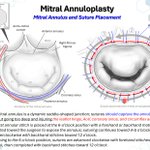
Mitral Annuloplasty—Annular Anatomy and Suture Placement🍩
👉Annular sutures should securely capture the mitral annulus while avoiding injury to the leaflet hinge, aortic valve, circumflex artery, and coronary sinus.


What is the durability of annuloplasty in patients with atrial functional mitral regurgitation associated with atrial fibrillation? Researchers at @CleClinicHVTI say the long-term durability is excellent. Log in to #JTCVS to read: hubs.la/Q04BqgfD0
@AskDrMarc @NadiaBakirMD

Aortic Valve & Root Anatomy #3: Basal Attachments and Ring Geometry
👉The aortic annulus is a virtual ring connecting the basal leaflet attachments, distinct from the anatomical ventriculo-arterial junction.
👉The leaflet insertions form a crown-shaped configuration, ascending toward the commissures and crossing the anatomical VA junction.
👉The sinotubular junction, interleaflet triangles, membranous septum, and aorto-mitral curtain define key relationships of the aortic root.


Aortic Valve & Root Anatomy #2: Root Configuration and Surrounding Structures
👉The aortic root comprises the sinuses of Valsalva, commissures, interleaflet triangles, and sinotubular junction.
👉The root is anchored within the cardiac fibrous skeleton, with aorto-mitral continuity and proximity to the membranous septum.
👉The aortic root lies adjacent to the pulmonary root, RVOT, and septal tricuspid annulus, reflecting its central anatomical position.

Fundamental Principles of Hemodynamics
👉Hagen–Poiseuille’s law describes flow resistance (Q ∝ r⁴), while Bernoulli’s principle explains pressure–velocity relationships across stenotic lesions.
👉Reynolds number predicts flow turbulence, while Laplace’s law relates pressure, chamber radius, and wall thickness to myocardial wall stress.
xitter.cf/WithAScalpel/status/21…
xitter.cf/WithAScalpel/status/21…


Why can a tiny change in blood vessel diameter dramatically alter blood flow?
The Hagen–Poiseuille equation reveals the power of the fourth: double the vessel radius, and blood flow increases 16-fold, assuming all other factors remain constant!
A beautiful example of how physics explains vascular physiology — and why arterioles play such a crucial role in regulating tissue perfusion.

Fundamental Principles of Hemodynamics: Ohm’s Law & Laplace’s Law
👉Hemodynamic Ohm’s law defines flow as Q = ΔP/R : pressure gradient drives flow, while vascular resistance opposes it.
👉Laplace’s law relates wall stress as T = (P × r)/(2h) : stress rises with ↑P or ↑r and falls with ↑h.
👉Clinically, these principles frame SVR/PVR, ventricular pressure/volume overload, hypertrophy/dilation, etc


Fundamental Principles of Hemodynamics: Bernoulli, Poiseuille & Flow Regimes
👉Bernoulli’s principle links velocity and pressure: flow acceleration through stenosis lowers lateral pressure; Doppler uses ΔP ≈ 4V².
👉Hagen–Poiseuille’s law shows Q = πr⁴ΔP/(8μL), making vessel, shunt, conduit, or cannula radius a dominant determinant of flow.
👉Laminar flow is orderly; turbulence is favored by ↑velocity, ↑diameter, and ↓viscosity, increasing energy loss and abnormal wall shear stress.


Aortic Valve & Root Anatomy #1: Aortic Leaflet and Root Structures
👉The aortic valve has three semilunar leaflets: right coronary (RCC), left coronary (LCC), and non-coronary (NCC).
👉Each leaflet has a hinge line, body, free margin, lunula, and nodulus of Arantius, contributing to central coaptation.
👉The RCC and LCC are associated with the right and left sinuses of Valsalva, giving rise to the RCA and LCA, respectively.
