

🔬📚Pathologist devoted to my work during the day, light-hearted when the sun goes down丨Mom 👶🏻👧🏻丨 Arctophile🧸丨Opinions my own, not medical advice
born in Granada🏔️ (Spain)
Joined June 2018
- Tweets368
- Following1.3K
- Followers1.4K
- Likes3.9K





Beatriz Agredano🐻🐼🐻❄️ retweeted

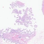
Well differentiated papillary mesothelial tumor
-Peritoneum
-Exophytic, papillary
-Bland, not invasive
-Mesothelial markers positive
-Don't call it mesothelioma
Dr. Mukhopadhyay CCMPS #lungpath #pathology #pathologists #pathtwitter #NIPPathX


Beatriz Agredano🐻🐼🐻❄️ retweeted

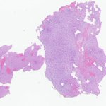
✅ Multinodular and vacuolating neuronal tumor (MVNT) 🎯:
• Discrete nodules in deep cortex and white matter
• Prominent cytoplasmic and pericellular vacuoles
• Neoplastic neurons show nuclear OLIG2 staining
• Driven by MAPK activation (MAP2K1 mutations)

ALT Discrete tumor nodules with vacuolar changes @IARCWHO

ALT Mature appearing neurons populate this nodule with vacuolar matrix changes @IARCWHO

ALT Neoplastic neuronal elements manifest nuclear OLIG2 immunoreactivity @IARCWHO

ALT Ramified CD34 immunoreactive neural elements are present @IARCWHO
¿Cómo os imagináis el infierno?🔥🔥🔥
Para mí es un lugar lleno de cilindros de próstata y de citologías vaginales ⚗️🧪🧫🧬🔬
#Pathology #PathologyFacts #Socorrito #SOS
Beatriz Agredano🐻🐼🐻❄️ retweeted

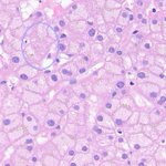
Male 35 💁🏻♂️
Radiology 🩻 : Huge liver mass suspecting Fibrolamellar carcinoma (FLC) 🤔
Pathology 🔬: Metastatic Adrenocortical Carcinoma (ACC) 😎
H&E ➡️ SF1 ➡️ SYN
Other markers:
CK focally ➕
CK7, HepPar 1, Arginase-1, HMB45, ERG, SALL4 ➖
INI1 Retained


Beatriz Agredano🐻🐼🐻❄️ retweeted

Cytomorphological features of acute myeloid leukaemia (AML) with t(8;21)(q22;q22.1);RUNX1::RUNX1T1
>>> Phenotypic clues that predict underlying cytogenetic/genetic abnormalities in myeloid malignancies: A contemporary review onlinelibrary.wiley.com/doi/… #AML #leusm

Beatriz Agredano🐻🐼🐻❄️ retweeted
Phenotypic clues that predict underlying cytogenetic/genetic abnormalities in myeloid malignancies: A contemporary review - Stone - 2023 - Cytopathology - Wiley Online Library onlinelibrary.wiley.com/doi/… #AML #leusm

Beatriz Agredano🐻🐼🐻❄️ retweeted
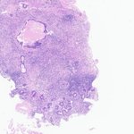
Histologic triad of Toxoplasma lymphadenitis includes
1⃣monocytoid B cell hyperplasia
2⃣reactive follicular hyperplasia
3⃣epithelioid histiocytosis
granulomas, multinucleate giant cells, and necrosis are absent
a similar triad occurs in CMV- & HIM-associated LAD
#HemePath #PathTwitter #Pathology
📽️on Reactive Lymphoid Hyperplasia
society-for-hematopathology.…
🙏@SocforHemepath
✅all of the above

Beatriz Agredano🐻🐼🐻❄️ retweeted
Crowded Gleason pattern 3
Glands are back-to-back and can look cribriform
Pitfall: calling it pattern 4
A keratin stain will highlight the architecture and show they are individual glands
Dr. McKenney. Prostatic Adenocarcinoma Grading: 2026 Update #USCAP #pathology #PathX

Beatriz Agredano🐻🐼🐻❄️ retweeted

30 yo woman with a complete hydatidiform mole one month ago. Endometrial aspiration revealed retained placental tissue. Is this atypical trophoblastic proliferation sufficient to diagnose choriocarcinoma? Opinions, please. #gynpath


Is it just me, or is diagnosing cholesterolosis and cholesterol polyps in a gallbladder weirdly satisfying?🤭
#path #pathology #PathTwitter


Astrocytoma, IDH-mutant, grade 2 🧠
📍Pending CDKN2A/B testing to determine if it will be molecularly reclassified to a higher grade.
IHC shows strong cytoplasmic and nuclear positivity for IDH1 R132H, along with a loss of ATRX nuclear expression in the tumor cells.
#neuropath🔬


Mantle cell lymphoma in a bone marrow biopsy. I love it when things work out well!
#pathology #hemepath👩🔬🩸


Beatriz Agredano🐻🐼🐻❄️ retweeted

A 9-year-old with headache, visual impairment, and endocrine deficits has a predominantly cystic, calcified suprasellar mass.
Diagnosis?
WHO clues:
• Mixed solid–cystic squamous epithelial tumour
• Stellate reticulum and wet keratin
• Hypothalamic–pituitary axis
• Activating CTNNB1 mutations
• “90% rule”: cystic, calcified, and enhancing cyst walls
• “Machinery oil” cyst fluid
• CNS WHO grade 1
Bonus: What is the arrowed inclusion, and which brain tumour is it classically associated with?
#Neuropathology #Pathology #MedEd #BrainTumor #Pathagonia


Beatriz Agredano🐻🐼🐻❄️ retweeted

#cytology #cytopath Clue of Mesothelioma vs Adenoca. In effusions:
♦️Groupings of mesothelial cells have rounded borders and look like the petals of a drawn flower
♦️Groupings of Adenocarcinoma cells often lack rounded bulbous individual cell contour and have a community border

Beatriz Agredano🐻🐼🐻❄️ retweeted

Morphologic Changes in Myocardial Infarction: A Microscopic Journey
Illustrating histopathology is one of the best ways to teach and learn! If you're interested in drawing, try illustrating histological changes—it helps in retaining concepts better and makes learning fun.
This image depicts the progressive changes in myocardial infarction, from the earliest cellular changes to fibrosis and scar formation.
Have tried illustration to simplify concepts for medical students. May not be comprehensive for speciality experts..
Suggestions and feedback welcome for improvements
@smlungpathguy @JMGardnerMD @DrGeeONE @pembeoltulu @adi_agnihotri @SaranyaS
@archibhat3 @vi_monappa @erikanuradha
@kriyer68 @DrAhmedKalebi
@PanditApoorva @RamyaPathology @DrNithyashree
@DrVasudevPrabhu @GIJamesMD @HermelinMD
@faheema_hasan @TristanRutland7 @KMirza
@pathridle_susan @DraEosina @cebulka26
@Baskotacytopath @Path_Matt @ariella8
@sranna86 @Thejaspatho @tumashankar
Check out my website and YouTube channel for more pathology illustrations, Medical School pathology educational content!
Website:ilovepathology.com/
YouTube: youtube.com/@ilovepathologyV…
Have you tried sketching histopathology slides? Let me know in the comments!
#Pathology #medicalschoolpathology #MedTwitter #medschool #MedicalEducation #Histopathology #Cardiology #MedicalIllustration #MedicalArt #LearningThroughDrawing #MyocardialInfarction #PathArt

Beatriz Agredano🐻🐼🐻❄️ retweeted

The remaining #Wallhangers . . . these pertain to tumor with "pink cells." (2/2)


Beatriz Agredano🐻🐼🐻❄️ retweeted
Oral epithelial dysplasia
WHO-2022: 3-tiered grading system
Defining dysplasia grade only in this manner oversimplifies the complexity of grading
Cytological atypia confined to the basal third may be sufficient for a diagnosis of severe dysplasia, depending on individual features present, particularly bulbous rete processes, budding and disorganization of basal cells, and marked pleomorphism
Pics Credit: Dr. Hernandez-Prera #USCAP25 #pathology #PathX


