

Open minded..easy going..
- Tweets146
- Following156
- Followers123
- Likes350



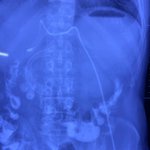
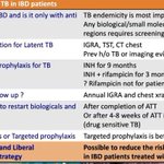


ALT a medical information about a patient


ALT The visual endoscopic retrograde appendicitis therapy procedure. (a) The colonoscopy was inserted into the cecum revealing mucosal hyperemia and bulging of the appendiceal orifice coated with a white moss-like substance. (b) A single-use video scope (blue arrow) was inserted into the appendiceal lumen along the guidewire through the appendiceal orifice by the working channel of the colonoscope. Massive pus was discovered during saline irrigation. (c) In one patient, 2 appendicoliths (red arrows) were identified in the appendiceal lumen by the single-use video scope after irrigation with saline solution. (d) Appendicoliths were extracted by a basket catheter under direct visualization of the single-use video scope. (e) The extraction of the appendicoliths was confirmed by the colonoscopy. (f) The appendiceal lumen was re-examined by the single-use video scope for signs of residual appendicoliths, ulceration, or appendicular stenosis. (g) In another patient without appendicolith, append


