

biotech / tech
New York, NY
Joined April 2026
- Tweets1.4K
- Following168
- Followers389
- Likes3.4K




Pinned Tweet
New hair loss treatment upgrade: Minoxidil Sulfate in a hydrogel-emulsion.
These images are from our Chinese pharmacy partner. They worked on products like L’Oreal’s Stemoxydine at one point.
#hairloss #hairgrowth #alopecia #biohacking
Preorder link:
folligenz.com/products/sulfo…
(global shipping)


🚨 Hair Loss Research Update 🚨
A man can have “female pattern” hair loss.
The hairline can stay in place while the hair behind it becomes thinner.
Chen et al. studied 38 Chinese Han patients with this pattern: 28 women and 10 men. They also examined 35 people without hair loss.
Under magnification, the patients had fewer hairs, finer hairs and more follicular units containing just one hair.
What caught my attention: the sides and BACK of the scalp could be involved too. Not just the crown.
That matters when assessing a possible transplant donor area.
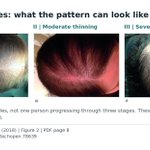
These are the paper’s actual scalp photographs. The man is on the left; the woman is on the right. Not before and after photos. 🔬🧵
Chen et al. (2021):
doi.org/10.3389/fmed.2021.64…
#HairLoss #Trichoscopy #HairScience #peptides #androgeneticalopecia #looksmaxxing #pharma #biohacker #Research

5/6 Early changes showed up in hair thickness, not just hair counts.
In the mildest Ludwig group, the crown already had significantly thinner hairs and a higher proportion of fine vellus hairs than controls.
The graphs compare 19 people at Ludwig I, 13 at II and 6 at III.
With greater severity, the overall pattern was lower density and thinner hairs, alongside more fine hairs and more units containing only one hair. Changes at the sides and back were clearer in the more advanced groups.
Read the colors as severity groups, NOT months of follow up. These are different people.
This study shows why “my hairline has not moved” is only one part of the picture.
doi.org/10.3389/fmed.2021.64…
#HairLoss #Trichoscopy #HairScience

6/6 Thinning describes what you see. It does not identify the cause by itself.
The evaluation chapter shows three separate examples here: a healthy scalp, telogen effluvium, and female pattern hair loss.
The chapter combines medical history, scalp examination and trichoscopy. A biopsy is sometimes needed, not automatically required for everyone.
Chen’s study specifically examined female pattern hair loss and excluded telogen effluvium and other scalp disorders. It also included only 10 affected men, so the findings should not be generalized to everyone.
Even BASP, Ludwig and similar labels do not fully describe what is happening at the sides and back, a limitation the authors highlighted.
My takeaway: do not stop at the hairline. Look at the whole scalp.
Chen et al.:
doi.org/10.3389/fmed.2021.64…
Photo source, Orasan et al.:
doi.org/10.5772/intechopen.7…
#HairLoss #ScalpHealth #Dermatology

🚨 Hair Transplant Research Update 🚨
Does your Norwood stage affect how many transplanted grafts grow?
A 2026 report from Vera Clinic looked at 386 men after FUE hair transplantation. At 12 months, its table reported average graft survival of:
94.3% for Norwood III.
88.4% for Norwood VII.
Interesting, but not proof that advanced baldness itself causes grafts to fail. This was one clinic, and the analysis did not separate the effect of hair loss severity from age.
The paper also has reporting inconsistencies, so I would treat these as preliminary figures, not personal predictions.
To understand the comparison, we need to look at the patterns. Norwood describes the overall balding pattern. BASP adds something useful: it separates hairline shape from thinning behind it. 🧵
Study:
doi.org/10.38124/ijisrt/26au…
Scalp photo source, Lee et al.:
doi.org/10.1016/j.jaad.2006.…
#HairLoss #HairTransplant #BASP
Tweet 2
2/6 First, what do those Norwood numbers actually mean?
III: pronounced recession at the temples.
III Vertex: the crown is the main area of loss.
IV and V: frontal and crown loss, with a bridge of hair still separating them. In V, that bridge is narrower and thinner.
VI: the bridge is gone and the two areas have joined.
VII: mainly a narrow horseshoe of hair remains around the sides and back.
These are descriptions of a pattern, not a countdown that everyone must follow.
The real scalp photographs here are labeled II and VI in their source. They are separate clinical examples, not before and after pictures and not patients from the transplant study.
Norwood chart and explanations, Gupta and Mysore:
doi.org/10.4103/0974-2077.17…
Scalp photographs, Orasan et al.:
doi.org/10.5772/intechopen.7…
#HairLoss #NorwoodScale #HairTransplant #pharma #biotech #peptides #research #Science #dermatology

5/6 Your hairline can stay in place while the hair behind it gets thinner.
Look at these two people from the BASP paper. The man and the woman were both classified LF2.
L means no hairline recession.
F2 means moderate diffuse thinning over the top.
That is why looking only at the front can miss part of the picture.
And F does NOT mean female. In this clinic sample, 749 of 1,768 men, or 42.4%, had an F pattern.
That is a finding in patients seeking care, not the percentage of all men with this pattern.
BASP can describe both men and women. But the graft survival figures we started with came only from men, and the transplant study did not calculate survival by BASP type.
BASP study:
doi.org/10.1016/j.jaad.2006.…
Graft study:
doi.org/10.38124/ijisrt/26au…
#BASP #HairLoss #HairScience
Tweet 6
6/6 So does this mean getting a transplant earlier guarantees better survival?
No. That question was not tested.
In the graft study, average age increased from 32.1 in Norwood III to 45.9 in VII. Age was not adjusted for. Another 42 patients did not complete the 12 month assessment and were excluded.
The paper’s chart also does not fully match its table. The exact figures need clarification before being treated as benchmarks.
My takeaway: use the pattern to describe the problem, not to calculate someone’s outcome from one number.
The evaluation chapter also emphasizes examining the scalp, assessing donor hair and documenting consistent photographic views. Norwood or BASP cannot replace that work.
The six photographs here show ONE person from different angles, not six stages of hair loss.
Graft study:
doi.org/10.38124/ijisrt/26au…
Evaluation chapter:
doi.org/10.5772/intechopen.7…
Photo source:
doi.org/10.1016/j.jaad.2006.…
#HairTransplant #HairLoss #Dermatology

🚨 Hair Loss Research 🚨
TWO YEARS of conventional treatment. Only modest improvement.
FOUR MONTHS after starting bicalutamide. Visibly better scalp coverage.
Then the side effects forced him to stop. ⚠️
Martínez Coronado et al. (2026) reported a 45 year old man with 15 years of hair loss and Norwood stage VI.
His previous treatments included topical minoxidil 5%, oral finasteride 1.25 mg/day and oral minoxidil 5 mg/day.
Both topical and oral minoxidil were stopped before bicalutamide 25 mg/day was started. Four months later, the authors documented visible regrowth, particularly around the crown.
That is what caught my attention: a visible response after years of hair loss and limited improvement with earlier treatment.
But this is ONE patient, assessed through photographs rather than standardized hair counts. It cannot prove that bicalutamide caused all the improvement or outperformed the previous drugs.
The adverse effects are central to this story, not a footnote. 🧵
doi.org/10.7759/cureus.11217…
#alopecia
#HairLoss #Bicalutamide #Dermatology #biohacker #pharma #peptides #Peptide #mtf #mtftransformation

2/3 Was this androgen blockade, or some unusual growth stimulating effect?
My read is that androgen receptor blockade is the more plausible explanation, rather than a special anabolic effect on hair.
Finasteride reduces the conversion of testosterone into DHT.
Bicalutamide acts at the receptor itself, interfering with signaling from both testosterone and DHT. Same hormonal system, different point of intervention.
The Masiello paper I mentioned adds an interesting detail. In prostate cancer models, the receptor could still reach the nucleus and bind DNA with bicalutamide attached. But it failed to recruit the helper proteins needed to switch the androgen responsive genes on.
Reaching the DNA is not the same as activating the genes.
That supports an antagonist mechanism, not proof of a SARM like hair growth effect. It was not a hair follicle experiment, and the case report did not measure hormone levels.
I still wonder: why no reported dutasteride trial before this? The paper does not explain. We cannot know whether it would have produced a similar response.
Case report:
doi.org/10.7759/cureus.11217…
Masiello et al. (2002):
doi.org/10.1074/jbc.M2033102…
#HairLoss #AndrogenReceptor #HairScience


3/3 The regrowth was interesting. The tradeoff was substantial. ⚠️
During treatment, he developed breast enlargement, breast pain, reduced libido and erectile dysfunction.
His liver enzymes also increased:
AST: 27 to 56 U/L.
ALT: 33 to 78 U/L.
The authors described these as mild elevations. Other possible causes were not fully investigated, so they could not attribute the blood test changes solely to bicalutamide.
The drug was stopped. Seven weeks later, the breast and sexual symptoms had reportedly resolved. Repeat liver tests were unavailable, so liver enzyme recovery was not confirmed.
Here is the other detail that interests me: he retained some improvement after returning to oral minoxidil 5 mg/day and a TOPICAL finasteride plus minoxidil formulation.
The paper does not say he resumed oral finasteride, and it does not specify how long the retained hair benefit lasted.
Could regrowth be induced and then maintained with a better tolerated approach? That is a research question, not something this single case establishes.
The authors explicitly caution against using this report to support oral bicalutamide for male pattern hair loss.
I wonder if he would have gotten these results with oral dutasteride. And if he could maintain the Bica results with dut as well 🧐🧬🥼🧪⚗️
doi.org/10.7759/cureus.11217…
#HairLoss #Bicalutamide #DrugSafety


5/7 The chronic telogen effluvium samples told another story.
They contained more follicles in telogen, the resting phase, but retained a terminal to vellus ratio of 7.1:1, which the paper described as normal.
Compare that with the 3:1 ratio reported in the pattern hair loss group.
In plain English: a shift toward more resting follicles is not the same finding as a shift toward miniaturized follicles.
The black arrows in the original image identify telogen follicular units.
Only THREE women had this diagnosis, so this is a very small comparison group.
doi.org/10.7860/JCDR/2026/88…
#TelogenEffluvium #HairLoss #HairScience

6/7 Blood tests added another piece to the picture.
18 of 50 women, or 36%, had ferritin below 40 ng/mL. Ferritin is used to assess iron stores, and 40 was this paper’s chosen threshold.
21 of 50 women, or 42%, had TSH above 4.2 mIU/L. TSH is a thyroid related blood test.
The authors classified 20 cases as subclinical hypothyroidism and one as overt hypothyroidism.
These findings could coexist with the underlying hair loss diagnosis. An abnormal blood test does not, by itself, explain everything happening inside a follicle.
The study did not establish that correcting these results would restore hair.
doi.org/10.7860/JCDR/2026/88…
#HairLoss #Ferritin #Thyroid


7/7 The takeaway is about getting the diagnosis right, not discovering a new cure.
This study combined scalp biopsies with blood tests because diffuse thinning can involve different follicle changes and accompanying biochemical abnormalities.
But neither ferritin nor TSH showed a statistically significant relationship with hair loss severity in this sample.
There was no healthy comparison group and no follow up measuring regrowth after iron supplementation or thyroid correction.
It was a small study at one hospital. Published in 2026, it used data collected in 2016 and 2017.
doi.org/10.7860/JCDR/2026/88…
#HairLoss #ScalpBiopsy #Dermatology

Hair Loss Research 🧵
Thinning all over your scalp does NOT automatically mean pattern hair loss.
In a scalp biopsy study of 50 women aged 15 to 45, researchers identified THREE different diagnoses:
28 women: diffuse alopecia areata, an immune related form of hair loss.
19 women: female pattern hair loss.
3 women: chronic telogen effluvium.
More than half had diffuse alopecia areata. But that is the finding in this hospital sample, not an estimate for all women with thinning hair.
The interesting part is what the biopsies actually showed: inflammation, miniaturization and changes in the hair cycle. Let’s look closer. 🔬🧵
Navya et al. (2026):
doi.org/10.7860/JCDR/2026/88…
#HairLoss #ScalpBiopsy #Dermatology #peptides #pharma
Accompanying image: Three original microscopy examples from Figures 1, 2 and 3, PDF page 2, organized alongside the reported diagnostic counts. These represent different conditions and magnifications, not changes before and after treatment. The diagnosis breakdown was 56% diffuse alopecia areata, 38% female pattern hair loss and 6% chronic telogen effluvium.

4/7 One finding deserves particular attention: fibrosis around follicles.
The authors reported perifollicular fibrosis in 15 of the 19 female pattern hair loss cases, about 79%.
“Perifollicular” means around the follicle. “Fibrosis” means fibrous tissue formation.
Panel c is their example of a miniaturized follicle with surrounding fibrosis.
Crucially, the study excluded scarring alopecia and still classified these cases as female pattern hair loss. It did not diagnose LPP in these women.
So this is a microscopic finding in a small subgroup, not evidence that 79% of women with pattern hair loss have LPP.
doi.org/10.7860/JCDR/2026/88…
#HairLoss #Fibrosis #Dermatology
An enlarged crop of original Figure 1c, which the authors identify as a miniaturized follicle with perifollicular fibrosis. The study reports 78.9% in its FPHL subgroup, while explicitly excluding patients with scarring alopecia.

3/7 Pattern hair loss had a different microscopic signature: MINIATURIZATION.
The follicles become smaller and produce finer hairs.
The authors found miniaturization in 16 of the 19 women classified as having female pattern hair loss, about 84%.
Their samples had a mean terminal to vellus ratio of 3:1: roughly three larger terminal follicles for every one fine vellus follicle.
In panel a, the original arrows identify terminal follicles. The triangular arrowheads identify vellus hairs.
The important clue is not just how many hairs remain. It is the mixture of larger and smaller follicles.
doi.org/10.7860/JCDR/2026/88…
#HairLoss #FemalePatternHairLoss #ScalpBiopsy
Original Figure 1a to 1c, PDF page 2, with the paper’s arrows and panel letters preserved. Panels a and b show variation in follicle size and miniaturized follicles. Panel c shows the fibrosis finding discussed next. The reported miniaturization frequency was 16 of 19 cases, or 84.2%.

2/7 The immune clue was hiding around the hair bulb.
Among the 28 women diagnosed with diffuse alopecia areata, 23 had lymphocytes clustered around the bulb, the lower part of the follicle.
That is about 82% of this diagnosis group.
Lymphocytes are immune cells. The authors describe their arrangement as a “swarm of bees.”
Look at panel b: the dark purple cells gather around the follicle’s lower portion.
This is why diffuse thinning can need a closer look. The visible complaint may be shedding, while the biopsy reveals an inflammatory process.
doi.org/10.7860/JCDR/2026/88…
#AlopeciaAreata #HairLoss #HairScience
Original Figure 2a and 2b, PDF page 2. Panel a is a cross section that the authors describe as showing inflammation and fibrosis around follicles. Panel b is a lengthwise section showing the lymphocytes around the bulb. Both are labeled H&E, ×40 in the paper.
