

Clinical Radiologist from Egypt interested in Chest Imaging and trying to make it interesting to others everywhere
Kuwait
Joined February 2011
- Tweets4.9K
- Following11
- Followers41.8K
- Likes3.7K







Pinned Tweet
من دواعي سروري كطبيب مصري أن أكون قد ساهمت بكتاب من تأليف فردي في المكتبة الطبية الدولية.
بعنوان «Challenging Cases in Interstitial Lung Diseases: A High-Resolution CT Approach» (CRC Press 2025)، يقدم الكتاب نهجاً عملياً منظماً لتشخيص أمراض الرئة الخلالية (التليف الرئوي) بدقة ومبكراً، آملاً أن يفيد الأطباء حول العالم و أن يساهم هذا العمل في تحسين رعاية هؤلاء المرضى وإدارة حالاتهم، مما ينعكس إيجاباً على حياتهم وتوقعاتهم المرضية.. أشكر كل من ساهم في إخراج هذا الكتاب إلى النور.


🔎 HRCT CHALLENGE: How many interstitial pneumonia patterns can you identify?
👩 Patient: 37-year-old woman
Sometimes, one pattern is not enough to describe the complexity of an HRCT examination.
🎥 Watch the video carefully and assess the distribution of fibrosis, ground-glass opacities, consolidation and pleural abnormalities.
💬 How many patterns can you identify? Which features support your interpretation?
I will discuss this case during Part 2 of my live lecture on the 2025 ERS/ATS Update on the Classification of Interstitial Pneumonias, using practical HRCT cases to explore combined patterns and their diagnostic implications.
📅 Friday, 30 October 2026
🕔 5:00 PM Cairo time
🎥 Missed Part 1? The recording is available, covering BIP, DAD, AMP and NSIP, with practical HRCT interpretation, diagnostic reasoning, and illustrated video cases.
📲 For registration and access details: WhatsApp +20 120 467 8455
#Radiology #HRCT #InterstitialLungDisease #ChestImaging #ERSATS #ThoracicRadiology
🔎 HRCT CHALLENGE: Can you identify the predominant interstitial pneumonia pattern?
A 70-year-old woman presents with shortness of breath. She has a history of breast cancer treated with bilateral mastectomy and breast reconstruction.
HRCT findings suggest an interstitial lung injury. The subsequent clinical course showed partial improvement after treatment, but the lung injury progressed to fibrosis.
What is the predominant HRCT pattern?
A️⃣ Bronchiolocentric interstitial pneumonia (BIP)
B️⃣ Diffuse alveolar damage (DAD)
C️⃣ Non-specific interstitial pneumonia (NSIP)
D️⃣ Organising pneumonia (OP)
💬 What is your answer, and which HRCT features support it?
I will discuss this case, the diagnostic reasoning and the relevant imaging clues during Part 2 of my live lecture on the 2025 ERS/ATS Update on the Classification of Interstitial Pneumonias.
📅 Friday, 30 October 2026
🕔 5:00 PM Cairo time
The session will focus on practical HRCT interpretation, overlapping patterns and the distinction between morphological patterns and their underlying causes.
📲 For registration and access details, contact me on WhatsApp: +20 120 467 8455
#Radiology #HRCT #ChestImaging #InterstitialLungDisease #ERSATS
📢 2025 ERS/ATS Update on the Classification of Interstitial Pneumonias
🔬 Part 2 — Live Online Lecture | Friday, 30 October
What has changed in the classification of interstitial pneumonias, and how can we translate these changes into better everyday HRCT interpretation?
Following our first session, I invite you to join me next Friday, 30 October 2026, at 5:00 PM Cairo time, for Part 2 of this practical, case-based lecture.
🔎 Part 1: More than a classification update
The Part 1 recording is available and includes:
▪️ Why was an update needed?
▪️ Four major advances and what has changed since 2013
▪️ Why separate interstitial and alveolar filling disorders?
▪️ BIP: The most important new pattern — essentials, practical reporting and the distinction between BIP and ACIF
▪️ From AIP to DAD: understanding idiopathic and secondary forms
▪️ DIP becomes AMP: what the new terminology means
▪️ NSIP: essentials from the 2025 ERS/ATS statement
▪️ MinIP and mosaic attenuation: technical considerations for HRCT interpretation
The session includes illustrated and video cases, practical radiological tips, pattern differentiation and diagnostic reasoning.
🎯 Part 2: What will we cover?
▪️ Organising pneumonia (OP)
▪️ Usual interstitial pneumonia (UIP)
▪️ Pleuroparenchymal fibroelastosis (PPFE)
▪️ Lymphoid interstitial pneumonia (LIP)
▪️ Respiratory bronchiolitis–interstitial lung disease (RB-ILD)
▪️ Combined interstitial pneumonia patterns
💡 What makes this approach different?
This is not simply a review of disease definitions or a reproduction of classification tables. We focus on how to interpret HRCT findings, distinguish overlapping patterns and use imaging to guide diagnostic reasoning.
Through practical examples, we explore questions such as:
🔹 When does a morphological pattern suggest a specific disease—and when does it not?
🔹 How can distribution and associated findings refine the differential diagnosis?
🔹 How should we approach BIP without automatically diagnosing HP?
🔹 How can we distinguish coexisting patterns and report them accurately?
🔹 How do we integrate HRCT, clinical history and histopathology?
The goal is to make the 2025 ERS/ATS classification practical, understandable and applicable to daily radiological practice.
📅 Join Part 2 live
Date: Friday, 30 October 2026
Time: 5:00 PM Cairo time
Format: Live online lecture
🎥 Missed Part 1? The recording is available, so you can catch up before the next session.
📲 For registration and access details, contact me on WhatsApp: +20 120 467 8455
I look forward to welcoming colleagues interested in a more practical and structured approach to interstitial lung disease and HRCT interpretation.
#Radiology #ChestImaging #HRCT #InterstitialLungDisease #ILD #ERSATS #ThoracicRadiology

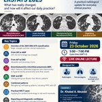
📢 SPECIAL LIVE ONLINE LECTURE
Updated Classification of Interstitial Pneumonias — ERS/ATS 2025
What has really changed, and how will it affect our daily practice?
The 2025 ERS/ATS statement introduces important changes to the international classification of interstitial pneumonias. Understanding these changes is essential for accurate HRCT pattern recognition, diagnostic reasoning and multidisciplinary discussion.
Join me for a 2-hour, clinically oriented session exploring the new classification through practical radiological examples.
🔎 What will we discuss?
▪️ From DIP to AMP: Why the terminology has changed.
▪️ From AIP to DAD: Understanding idiopathic and secondary forms.
▪️ Bronchiolocentric interstitial pneumonia (BIP): A new pattern, its HRCT features and its relationship to hypersensitivity pneumonitis and other causes.
▪️ NSIP and BIP: Distinguishing non-fibrotic from fibrotic patterns.
▪️ Combined patterns: Recognising overlapping patterns, including PPFE with UIP.
▪️ Practical HRCT cases: Applying the updated terminology to real-world imaging findings.
▪️ Diagnostic confidence: What should we report, and what still requires clinical and multidisciplinary integration?
🎯 Designed for: Radiologists, pulmonologists and physicians involved in the diagnosis and management of interstitial lung diseases.
📅 Friday, 23 October 2026
🕔 5:00–7:00 PM Cairo time
💻 Live online lecture
📩 To join or enquire about participation, contact me via WhatsApp:
+20 120 678 4555
A focused opportunity to understand the new classification and translate its changes into everyday clinical and radiological practice.
I look forward to meeting you online!
#InterstitialLungDisease #ILD #ThoracicRadiology #HRCT #ERS #ATS2025

Dr Khaled Ahmed Abuzeid retweeted
SPECIAL LECTURE
Updated Classification of Interstitial Pneumonias — ERS/ATS 2025
Before we continue with the remaining HRCT pattern sessions of the course — Consolidation, Cystic and Mosaic Patterns — I will give a special lecture on the Updated ERS/ATS Classification of Interstitial Pneumonias (2025).
📅 Friday, 23 October 2026
⏰ 5:00–7:00 PM Cairo time
💻 Live Online Lecture
The lecture will focus on the important changes in the updated classification and, most importantly, how these changes can be applied in everyday HRCT practice.
🔹 What is new in the 2025 classification?
🔹 What has changed from previous classifications?
🔹 How should we approach these entities on HRCT?
🔹 What are the practical implications for radiologists?
Interested in joining?
Please contact me on WhatsApp:
📱 +20 120 678 4555
The remaining HRCT pattern sessions will continue after this special lecture.
🤖 Made with AI

Some players play football. Others inspire generations.
Mohamed Salah and Cristiano Ronaldo have given us countless moments of joy, passion and inspiration. They have made us believe, dream and love the game even more.
Legends deserve respect. Always. ❤️⚽


SPECIAL LECTURE
Updated Classification of Interstitial Pneumonias — ERS/ATS 2025
Before we continue with the remaining HRCT pattern sessions of the course — Consolidation, Cystic and Mosaic Patterns — I will give a special lecture on the Updated ERS/ATS Classification of Interstitial Pneumonias (2025).
📅 Friday, 23 October 2026
⏰ 5:00–7:00 PM Cairo time
💻 Live Online Lecture
The lecture will focus on the important changes in the updated classification and, most importantly, how these changes can be applied in everyday HRCT practice.
🔹 What is new in the 2025 classification?
🔹 What has changed from previous classifications?
🔹 How should we approach these entities on HRCT?
🔹 What are the practical implications for radiologists?
Interested in joining?
Please contact me on WhatsApp:
📱 +20 120 678 4555
The remaining HRCT pattern sessions will continue after this special lecture.
🤖 Made with AI
Honored to have been invited to participate in the First Syrian Radiology Conference – “Updates in Radiology” in Hama, and truly grateful for the warm welcome and wonderful hospitality.
It was a pleasure to share two lectures on fibrotic ILD and HRCT and the updated ERS/ATS classification of interstitial pneumonias, and to have the opportunity to exchange knowledge and experiences with colleagues.
My sincere thanks to the Syrian Radiology Association, Hama Medical Syndicate, and the organizing committee for the kind invitation and excellent organization.
A memorable experience, and I hope this will be the beginning of many more opportunities to connect and collaborate with colleagues in Syria.


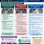
DLD COURSE — DIFFUSE LUNG DISEASES
Pattern-Based HRCT Approach
The DLD Course is continuing with a practical, pattern-based approach to HRCT interpretation, focusing on how to recognize the CT pattern, narrow the differential diagnosis, and integrate the findings into a confident radiologic diagnosis.
🎥 Recorded sessions currently available:
🔹 Reticular Pattern — 1 hr 50 min
🔹 Nodular Pattern — 5 hrs 7 min
🔹 Ground-Glass Pattern — 19 hrs 41 min
The poster shows the detailed topics covered within each pattern.
📚 The course is not finished yet!
The remaining sessions will continue from:
📅 Friday, 25 September 2026
with the upcoming patterns:
🔸 Consolidation Pattern
🔸 Cystic Pattern
🔸 Mosaic Pattern
How to participate?
You can choose either:
✅ Join the whole DLD Course and follow the remaining sessions
or
✅ Purchase selected recorded video lectures according to your interests.
📱 For registration and details, contact WhatsApp:
+20 120 678 4555
If you are interested in developing a structured and practical approach to diffuse lung disease on HRCT, you are welcome to join the course.
Let’s make HRCT interpretation more practical together.
— Khaled A. Abuzeid
Consultant Radiologist | Chest Imaging
🤖 Made with AI

Halfway there. 🔹
The first half of the course is now complete, with 13 sessions delivered. We are taking a short 3-week break before continuing on September 25.
For those who have been considering joining, enrollment is still open.
New participants will receive access to the completed sessions and course materials, giving them the opportunity to catch up during the break and join us for the second half from September 25.
The upcoming sessions will continue with a strong focus on practical approach, case-based learning, and structured HRCT interpretation, taking the concepts from pattern recognition to clinical diagnosis.
We resume on September 25.
If you are interested in joining, this is an ideal time to enroll and use the break to catch up with the first half.
Special academic group arrangements are available for:
— Radiology residents and fellows
— Pulmonary residents and fellows
— Radiology or Pulmonary departments and institutes
— Academic training groups
📲 For registration, academic group arrangements, and further information, contact us on WhatsApp:
+20 120 678 4555
🤖 Made with AI

Honored to be invited as a guest speaker at the 1st Scientific Conference of the Syrian Radiologists Association – “Updates in Radiology”, taking place in Hama, Syria, on September 18–19.
I will be presenting two lectures focused on interstitial lung disease and the role of HRCT in diagnosis:
• My Approach to Fibrotic Interstitial Lung Disease: From HRCT Pattern to Diagnosis
• The Updated ERS/ATS Classification of Interstitial Pneumonias: What Radiologists Need to Know
It is a great pleasure to contribute to this important scientific event and to be part of a program bringing together radiologists and experts from Syria and around the world.
Looking forward to meeting colleagues and sharing knowledge in Hama.
يسعدني ويشرّفني أن أكون محاضراً ضيفاً في المؤتمر العلمي الأول لرابطة الشعاعيين السوريين «المستجدات في علم الأشعة»، والذي سيُعقد في مدينة حماة – فندق أفاميا الشام، بتاريخ 18–19 أيلول.
سأشارك بمحاضرتين حول أمراض الرئة الخلالية ودور HRCT في الوصول إلى التشخيص:
• My Approach to Fibrotic Interstitial Lung Disease: From HRCT Pattern to Diagnosis
• The Updated ERS/ATS Classification of Interstitial Pneumonias: What Radiologists Need to Know
يشرفني أن أكون جزءاً من هذا الحدث العلمي المميز، الذي يجمع نخبة من أطباء الأشعة والخبراء من سورية ومن مختلف أنحاء العالم.
أتطلع للقاء الزملاء والمشاركة في هذا الحدث العلمي المميز في حماة.


A special moment at ERS Congress 2026 in Barcelona.
Delighted to see Challenging Cases in Interstitial Lung Diseases: A High-Resolution CT Approach displayed at the CRC Press / Taylor & Francis exhibition. Grateful to Shivangi Pramanik and the team for making this possible.
To my friends and colleagues attending ERS—enjoy the Congress! I hope you get a chance to spot the book. 📚
#ERS2026 #InterstitialLungDisease #HRCT #ThoracicRadiology #Pulmonology #Radiology

A challenging chest CT—and a perfect example of why pulmonary nodule interpretation matters.
A 65-year-old man with chest pain and a remote history of severe COVID-19 pneumonia requiring hospitalization 6 years ago.
The CT shows two very different stories:
🔹 Chronic interstitial lung abnormalities
• Upper-lobe predominant pleural/parenchymal fibroelastosis–like change (PPFE pattern)
• Lower-lobe predominant residual organizing-pneumonia changes
🔹 Multiple pulmonary nodules along the adenocarcinoma spectrum
• Dominant right upper lobe juxtapleural part-solid nodule, 23 mm, with a 21-mm solid component
• Left upper lobe peripheral solid nodule, 5 mm
• Left upper lobe para-aortic pure ground-glass nodule, 7 mm
The challenge is not simply to see the nodules.
It is to understand:
Which findings are related to the previous lung injury—and which findings require a completely different approach?
And, more importantly:
How should we interpret and manage incidentally detected subsolid nodules?
This is exactly the type of case discussed in my lecture:
“Adenocarcinoma: How to Interpret Incidentally Suspected Adenocarcinoma Spectrum”
We go through the Fleischner Society 2017 recommendations for incidentally detected pulmonary nodules, and also discuss how the approach changes when we are working within a lung cancer screening program using Lung-RADS v2022.
The same nodule can require a different framework depending on the clinical context.
🎓 The full DLD Course is currently available for enrollment.
The Adenocarcinoma lecture is also available for X subscribers.
If you interpret chest CTs, this is one lecture worth having in your toolbox.
📚 DLD Course — Halfway Completed!
We have now completed half of the DLD Course🎯
So far, we have covered:
🔹 Reticular pattern — 1 session
🔹 Nodular pattern — 3 sessions
🔹 Ground-glass pattern — 9 sessions
Each session is approximately 2 hours, with a strong focus on practical HRCT interpretation and clinical decision-making.
We have now completed the Ground-Glass Pattern, including the pulmonary adenocarcinoma spectrum.
⏸️ Planned 3-Week Break
We will take a planned break for three Fridays and resume the course on:
📅 Friday, September 25
We will then continue with:
🔸 Consolidation pattern
🔸 Cystic pattern
🔸 Mosaic pattern
New participants are welcome!
The 3-week break is a great opportunity to join the course now and catch up through the recorded sessions before the live sessions resume.
Half of the course completed — and we still have a lot more HRCT to explore!
🤖 Made with AI


GROUND-GLASS OPACITY PATTERN — PART 9
PULMONARY ADENOCARCINOMA
From GGO to Practical Reporting
What does the CT really tell you when you encounter a ground-glass or part-solid pulmonary nodule?
In this session, we will explore the pulmonary adenocarcinoma spectrum from a radiologic perspective:
🔹 AIS • MIA • LPA • IMA
🔹 Pure ground-glass nodules
🔹 Part-solid nodules
🔹 Consolidative and multifocal/diffuse presentations
🔹 Cystic airspaces associated with adenocarcinoma
🔹 Radiologic–pathologic correlation
🔹 Real-world cases and diagnostic pitfalls
But the most important part is the practical approach:
What should the radiologist actually report?
We will discuss reporting recommendations and how to approach these lesions in clinical practice, including:
🔸 When and how to apply the Fleischner guidelines
🔸 When Lung-RADS applies
🔸 How to describe pure GGO and part-solid nodules
🔸 How imaging features can suggest the underlying adenocarcinoma spectrum
🔸 What matters on follow-up and what should trigger further action
Friday, August 21, 2026 | 5:00 PM Cairo Time
💻 Live Online Session
Individual registration is available for those interested specifically in this topic
📲 Registration & course details:
WhatsApp: +20 120 678 4555
🤖 Made with AI

Next Session | Ground-Glass Opacity Pattern — Part 8
We have already discussed many of these diseases in previous pattern-based sessions—but this time, we will look at them from a completely different perspective.
The question is not simply “What diseases cause ground-glass opacity?”
It is:
What diseases can present with a chronic ground-glass pattern, and how can we recognize them when they don't show their more familiar appearance?
In this session, we will discuss:
🔹 Atypical sarcoidosis presenting with chronic GGO
Moving beyond the typical sarcoidosis pattern of perilymphatic nodules that we discussed in the Nodular Pattern session, and exploring the different HRCT patterns of chronic GGO-predominant sarcoidosis.
🔹 Respiratory bronchiolitis and follicular bronchiolitis revisited
Previously discussed as causes of centrilobular ground-glass nodules. This time, we will see cases presenting with patchy GGO rather than discrete centrilobular nodules, and explore their associations with smoking-related disease and connective tissue disease/LIP, respectively.
🔹 Pneumoconiosis presenting as chronic GGO
Not every pneumoconiosis presents with the familiar nodular pattern. We will look at less typical GGO-predominant presentations and the clues that help us recognize them.
🔹 Pulmonary alveolar proteinosis (PAP)
Beyond the classic crazy-paving appearance, with multiple real-world cases demonstrating the spectrum of HRCT appearances and potential diagnostic pitfalls.
🔹 Lipoid pneumonia
How can we recognize lipoid pneumonia when obvious fat attenuation is absent? We will integrate the radiological clues with the clinical history and discuss how to distinguish lipoid pneumonia from mucus bronchogram and mucus impaction.
This is the 8th session of our Ground-Glass Opacity Pattern series. After exploring both acute and chronic GGO presentations throughout the previous sessions, we are now continuing with the chronic GGO spectrum.
And there is still one more session to complete the GGO pattern:
Part 9 will move into adenocarcinoma and other lung cancers presenting with ground-glass opacity, completing the GGO pattern from inflammatory and interstitial diseases to neoplastic causes.
Diseases do not always read the textbook. Their HRCT appearance can change, and recognizing the pattern in front of you is the first step toward the correct diagnosis.
Diffuse Lung Disease HRCT Pattern-Based Course
📅 Friday, August 14, 2026
⏰ 5:00 PM Cairo Time
💻 Live online via Zoom
Case-based • Pattern-based • Practical HRCT interpretation
Previous session recordings are available to course participants.
🤖 Made with AI

Hemoptysis + diffuse ground-glass opacity = DAH? Not always.
A 65-year-old man presented with cough, breathing difficulty and hemoptysis.
His HRCT showed bilateral bat-wing ground-glass opacities, marked cardiomegaly and a dilated main pulmonary artery.
At first glance, hemoptysis with diffuse GGO and consolidation may make us think of diffuse alveolar hemorrhage (DAH), particularly from systemic vasculitis.
But there is an important diagnostic trap here:
Pulmonary edema can also cause hemoptysis.
So when you see diffuse GGO in a patient with hemoptysis, don't let the clinical symptom alone drive you toward DAH. Look at the distribution of the GGO and, importantly, the cardiovascular findings.
In this case, the bat-wing distribution, marked cardiomegaly and dilated pulmonary artery provide important clues toward pulmonary edema related to cardiac disease and pulmonary hypertension.
👉 Hemoptysis does not necessarily mean DAH.
Sometimes, the lung is telling you about the heart.
This is also one of the important pitfalls I discussed in my recent acute GGO session of the Diffuse Lung Disease HRCT course.