

Specializing in sports foot and ankle orthopedic surgery.
Nashville, TN
Joined December 2011
- Tweets499
- Following429
- Followers390
- Likes431
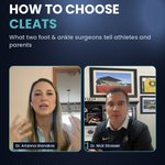



How to choose cleats: match the surface, get the fit right, and don't assume the most aggressive grip is safest. Linemen need a wider base; women's feet tend to be wider in front, narrower at the heel. From Athletes Corner with @AGianakosDO. #cleats


Is turf worse for your ankles? It's less about the surface and more about whether the foot can release while the body keeps rotating. New Athletes Corner episode with @AGianakosDO is on YouTube now. #sportsmedicine #turf
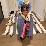
Happy Halloween! My daughter went headless this year, using the spine models I've saved since residency. They finally came in handy. 🦴🎃 Stay safe out there, and watch those ankles on dark porch steps.

ALT A girl in a headless costume with a model spine above the collar
How long does an Achilles rupture take? Plan on 6–12 months. One patient: "I returned to sport at about nine months. But I didn't feel like myself until 12." Full episode with @AGianakosDO on YouTube. #achilles
Achilles rupture recovery, realistically: motion at 2–3 weeks, out of the boot at 6–8, strengthening at ~12, back to sport at 6–12 months. And it's milestones, not just time. From Athletes Corner with @AGianakosDO. #achilles


Today's Achilles repairs and rehab often allow motion within 2–3 weeks. But no matter what you're having done, "biology is biology." New Athletes Corner episode with @AGianakosDO is on YouTube now. #achilles #sportsmedicine
Is a broken ankle just a bad high ankle sprain? Often it's the worst kind: the same ligaments tear, plus the bone. That's why fixing the fracture is only part of the job. Full episode with @AGianakosDO on YouTube. #highanklesprain
An ankle fracture is more than a broken bone. In a fracture-dislocation, 4 structures fail: the AITFL, the fibula, the PITFL and the deltoid side. Only one of them is bone. From Athletes Corner Ep. 1 with @AGianakosDO. #anklefracture


Why do ankles dislocate? The foot is stuck on the ground while the whole body rotates around it, and that force has to go somewhere. New Athletes Corner episode with @AGianakosDO is live on YouTube. #anklefracture #sportsmedicine
Your Achilles is the strongest tendon in your body — so what actually happens when it snaps? It usually tears a few centimeters above the heel, where the blood supply is weakest, and a lot of people can still walk afterward, so it gets missed. The quick test: lie face down and squeeze the calf. If the foot doesn't move, get it checked. Caught early, it heals well with surgery or in a boot.
Animation created with AI tools and reviewed by me for accuracy; voiceover is my own. Not medical advice.
#achillesrupture #achillestendon #sportsmedicine #footandankle #orthopedics #basketball #pickleball #anatomy #NickStrasserMD
A high ankle sprain isn't a normal sprain. It's the syndesmosis — the ligaments holding the tibia and fibula together — and every step pushes the bones apart. That's why it heals slowly and sometimes needs surgery.
#highanklesprain #syndesmosis #anklesprain #sportsmedicine #footandankle #orthopedics #football #hockey #anatomy #NickStrasserMD
Roll your ankle and this is the ligament that goes first: the ATFL. Roll harder and the CFL goes too. Here's what's actually happening inside the joint. Not medical advice — if your ankle keeps giving way, get it looked at.
#anklesprain #ATFL #sportsmedicine #footandankle #orthopedics #anatomy #athlete #rolledankle #sportsinjury #NickStrasserMD
So what did we do?
After several long discussions with the patient and his family, we elected to pursue joint preservation.
At 19 years old, none of the options were particularly simple. Ankle fusion was certainly a consideration. A bipolar osteochondral allograft was another. Ultimately, we elected to reconstruct the lateral talar surface with a hemitalar osteochondral allograft, combined with a closing-wedge tibial osteotomy to shift load away from the involved lateral side of the ankle.
We started through a direct anterior approach and identified the extent of the talar lesion. Preoperative CT was particularly helpful for planning the resection.
Kirschner wires were used to define the resection, and I've found a micro-reciprocating saw very useful for making these cuts. I also place a Chandler posteriorly to protect the back of the joint. A Hintermann-type K-wire distractor can provide additional visualization when needed.
Once the diseased talar segment was removed, the time-consuming part began: shaping the donor allograft to reproduce the defect as precisely as possible.
Once satisfied with the fit, we incorporated BMAC and tibial autograft and sealed the graft margins with fibrin glue.
Finally, we performed a relatively modest closing-wedge tibial osteotomy—not an aggressive correction, but enough to decrease loading across the reconstructed lateral ankle. This was stabilized with a medial plate.
There are obviously several ways to approach a problem like this, and the long-term durability of any joint-preservation strategy in a case this advanced remains an important question.
Would you have approached this differently?
Fusion? Bipolar allograft? A different osteotomy or reconstruction?
Open to feedback, comments—and criticism. These are the cases where I think the discussion is particularly valuable.
One of the best parts of AOFAS is the conversation—reconnecting with colleagues, hearing new ideas, reinforcing familiar ones, and making new friends.
Honored to have the opportunity to present on return to play in achilles tendinopathy, fifth metatarsal fractures and syndesmosis injuries.
Some of my favorite conversations happened behind the microphone for the AOFAS Orthopodcast, particularly while interviewing this year’s award winners. A few topics that stood out:
• Weight-bearing CT and syndesmotic instability—and the importance of considering the multiple planes in which instability can occur.
• Congruent versus incongruent ankle arthritis, and fixed versus mobile-bearing ankle replacements.
• The interosseous talocalcaneal - cervical ligament and its potential role in subtalar instability and subfibular impingement.
• Osteochondral lesions and emerging approaches that may help promote repair.
I came away with new questions, fresh perspectives, and plenty to think about as we work to help athletes and active people keep moving.
These episodes should be out soon. Subscribe to the @AOFAS Orthopodcast so you never miss a conversation!
This is an interesting case of a 19-year-old male who presented with 9 months of progressive ankle pain and deformity.
It started with what he describes as a relatively minor ankle sprain. He was able to walk on it and eventually returned to most of his normal activities, including a job that required him to spend extended periods of time on his feet.
But the ankle never really returned to normal.
Several months later, while on a trip that required extensive walking, he developed significant pain and swelling that became quite limiting. An MRI was obtained shortly afterward, and he eventually presented to us because of increasing pain, progressive deformity, and difficulty with everyday activities.
One other interesting part of his history: he previously had an osteochondral injury involving his knee that required fairly extensive surgical treatment.
He is otherwise healthy, currently in school, and wants to remain active. Laboratory workup prior to presentation was essentially normal.
In the video, I’ve included his X-rays, MRI, and CT scan.
For the foot and ankle surgeons out there:
What do you think is going on?
More importantly, in a 19-year-old with these findings, how would you approach treatment?
Curious to hear how others would think through this one.
There were a lot of great comments on this case, particularly about weight-bearing X-rays and CT.
At two weeks, the patient could put some weight on the ankle—but not enough for a true weight-bearing comparison. Based on the medial clear-space widening, lateral talar translation, and concern about the fibular reduction, I recommended early revision.
This is not a decision to take lightly. It means another operation, another recovery, and more risk. However, this was a young, active patient whose ankle was important for both sports and work. The decision might have been different for a lower-demand patient or someone with other risk factors.
Would CT have been reasonable? Absolutely. It could have better defined the fibular rotation, syndesmotic reduction, and articular injury.
Biomechanical studies show that external rotation of the fibula alters ankle contact mechanics, including increased posterolateral tibiotalar contact pressure. Does that prove he would have developed arthritis? No. The long-term clinical significance remains unclear. Anecdotally, however, this is a pattern in which I have seen early posterolateral arthritis develop.
Arthroscopy gave us more information. The fibula surprisingly appeared to be out to length, but it was externally rotated and the syndesmosis was malreduced. We also found articular fragments peeled from the medial talus and the deltoid ligament interposed in the medial gutter. Fortunately, the posterolateral tibial cartilage looked better than expected.
We revised the fibula, repaired the deltoid, and revised the syndesmosis. The fracture was still mobile, so no osteotomy was needed.
The article in the reel highlights useful clues on the mortise view: a pointed or blade-shaped fibula suggests external rotation, while a spoon-shaped profile suggests internal rotation.
Does this matter long term? How close is close enough? Would you revise early—or wait?
#AnkleFracture #FootAndAnkle #OrthopedicSurgery
I’m jumping on the trend from @ankl_man and drkevinmartin_ortho who have shared some interesting cases and asking how others would approach them.
This patient presented to me after undergoing ankle fracture fixation elsewhere. These first images show the original injury and the reduction obtained at surgery.
At first glance, the fixation may appear reasonable—but there are some subtle nuances in the fibular profile and overall ankle alignment that deserve a closer look.
Continue with immobilization and allow it to heal—or recommend early revision? WBAT in a boot? CT scan indicated?
This is meant to be a discussion about decision-making—not criticism. These cases are often much clearer in retrospect than they are in the operating room. #mondaymorningquarterback
What would you be watching for on the next set of X-rays?
#AnkleFracture #OrthopedicSurgery #FootAndAnkle #SportsMedicine #MedicalEducation #SurgicalDecisionMaking
The hardest part of Achilles recovery often isn’t getting the tendon to heal—it’s rebuilding the calf.
Calf muscle loss begins quickly after an Achilles rupture. Even when the splint comes off after only two weeks, the injured leg can already look and feel dramatically different.
That’s why the later phases of recovery are about more than tendon healing. They’re about restoring strength, power, and confidence.
One of the biggest things I see patients neglect is consistent, progressive calf strengthening. During the first year, that often means training the calf three or four times per week and gradually incorporating:
• Weighted calf raises
• Eccentric strengthening
• Seated calf raises
• Standing calf raises
Both seated and standing exercises matter because they emphasize different parts of the calf muscle complex.
A healed tendon does not automatically mean a fully recovered athlete. Rebuilding the calf requires progressive loading, consistency, and time.
For the physical therapists and athletic trainers out there: What are your favorite strategies for restoring calf strength after an Achilles rupture? Are there specific exercises, progressions, or benchmarks you find particularly helpful?
Watch the full video, “Achilles Rupture Rehab: What Recovery Really Looks Like,” on YouTube.
#AchillesRupture #AchillesRehab #CalfStrength #PhysicalTherapy #SportsMedicine #ReturnToSport
By weeks 6–12 after an Achilles rupture, most patients are beginning to walk more normally—but the tendon is still healing.
During this phase, we gradually progress toward full weight bearing, work on range of motion, and begin rebuilding muscle activation and strength. Blood-flow restriction training can be particularly useful because it allows us to challenge the muscle with lighter loads while continuing to protect the tendon.
But this is also a phase when patients can begin to feel more capable than the tendon actually is.
One of the biggest risks is an unexpected slip or misstep—especially getting in and out of the shower without the boot. I have seen patients reinjure the Achilles during these seemingly routine moments after both operative and nonoperative treatment.
The takeaway: increasing activity does not mean abandoning protection. Progress gradually, follow your rehabilitation plan, and remain especially careful during transitions when the boot is off.
Watch the full video, **“Achilles Rupture Rehab: What Recovery Really Looks Like,”** on YouTube for the complete recovery timeline.
#AchillesRupture #AchillesRehab #AchillesRecovery #BloodFlowRestriction #PhysicalTherapy #SportsMedicine #ReturnToSport
What does recovery actually look like after an Achilles tendon repair?
During the first two weeks, the priority isn’t running—or even aggressive rehabilitation. It’s protecting the repair, allowing the incision to heal, controlling swelling, and minimizing muscle loss.
From weeks two through six, rehabilitation becomes a careful balancing act. The Achilles benefits from early movement and controlled loading, but doing too much too soon can stretch the healing tendon and compromise its function.
This is where an experienced physical therapist can be invaluable. Rehabilitation may include:
• Protected weight bearing
• Carefully controlled range of motion
• Swelling management
• Blood-flow restriction training to help limit muscle atrophy
The goal is to introduce enough movement and stress to encourage recovery—without overwhelming the healing tendon.
Watch the full video, “Achilles Rupture Rehab: What Recovery Really Looks Like,” on YouTube for the complete rehabilitation timeline.
#AchillesRupture #AchillesRehab #AchillesRepair #BloodFlowRestriction #PhysicalTherapy #SportsMedicine #ReturnToSport