
The Society of Pediatric Neuroimaging | A dynamic faculty from the world’s leading children’s hospitals leading the way in education and research
Joined May 2019
- Tweets3.1K
- Following206
- Followers6.4K
- Likes3.4K




SPIN Module 2 • Day 2
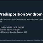
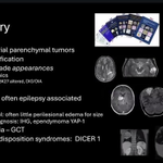
An insightful talk by @sanjayprabhu on Tumor Predisposition Syndromes!
From syndrome-specific screening protocols to reporting pitfalls, a practical approach to recognizing key imaging findings, avoiding missed lesions, and improving surveillance.


SPIN Module 2 • Day 2
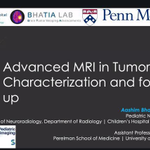
A fantastic talk by @draash on Advanced MRI in Tumor Characterization and Follow-up!
Key insights into the tumor microenvironment and advanced imaging biomarkers, emphasizing standardized protocols and their translation into clinical practice.

SPIN Module 2 • Day 2
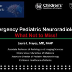
A fantastic talk on Emergency Pediatric Neuroradiology by @laura1127md.
From complications of sinusitis to subtle spine trauma findings and metabolic mimics, recognizing key imaging clues can make all the difference.
The takeaway? Look beyond the obvious!


SPIN Module 2 • Day 2
Mastering infratentorial pediatric brain tumors! A fantastic talk by Dr Eleonora Piccirilli highlighting the importance of a protocol-driven approach, recognizing classic and atypical imaging patterns, and integrating histology with molecular profiling.


SPIN Module 2 • Day 2
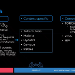
A stellar talk by @AmalSalehNour on a systematic approach to CNS infections, from etiology to clinical context!
A structured approach to imaging common bacterial and viral infections, with insights into distinguishing congenital from acquired infections.


SPIN Module 2• Day 2
A practical approach to MRI in pediatric acute demyelinating syndromes (ADS) with Dr Li-Tal Pratt!
From initial diagnostic imaging to recent changes in the diagnostic criteria, this talk explores all the key facets of ADS.


SPIN Module 2• Day 2
Dr Ankita Gupta delivered a stellar session on Infection-Triggered Encephalopathy Syndromes, covering all the important clinical and radiological aspects.
Timing of imaging and DWI are key factors in evaluation!


SPIN Module 2 • Day 2
Dr Jane McEniery is opening today’s program with an excellent session on Pediatric Supratentorial Tumors.
From imaging patterns and diffusion to PLNTY, DNET, MVNT, ganglioglioma, PXA, and molecular classification — a highly practical start to the day.

Join us for Module 2, Day 2 of the Society of Pediatric Neuroimaging Fellowship Teaching Program 2026/27 on Oct 24th!
Featuring expert-led sessions covering pediatric brain tumors, demyelinating disorders, CNS infections, emergency neuroradiology, advanced MRI, and more!

SPIN | Society of Pediatric Neuroimaging retweeted

Just when you thought you knew all the possible layers of intracranial hemorrhage…(yeah me too!)... neonatal subpial hematoma. See a nice thread from @spinacademics from 10/15/2025 for more information.


It’s #SPIN Tweetorial Wednesday!
An infant with temporal white matter edema, pallidal injury and strikingly tortuous intracranial arteries.
What connects these findings?
This week: Menkes disease. 🧵

Subdural collections can also occur.
In an infant with subdural collections, review the arteries and background brain carefully. The combination of tortuosity, abnormal myelination and parenchymal injury should raise suspicion for Menkes disease.
Two different diffusion patterns in the same examination:
• Pallidal lesions with restricted diffusion.
• Temporal white matter edema with facilitated diffusion.
The latter reflects vasogenic edema.
Look beyond the vessels.
MRI may show delayed myelination, cerebral volume loss and transient, sometimes asymmetric, temporal white matter edema.
Basal ganglia injury may coexist.
Menkes disease is an X-linked copper transport disorder caused by ATP7A variants.
An important imaging clue: diffuse intracranial arterial elongation and tortuosity, which may already be apparent in the neonatal period
Next on The Cortex Club × SPIN Physics Talk is an insightful session with Dr Haidar Almansour, as he explores Fast MR Scanning and Deep Learning-Based Reconstruction Methods
📅 October 28, 2026
🕒 10 AM EST | 3 PM GMT | 4 PM CET | 8:30 PM IST
🔗 Register: bit.ly/spinphysicssessions

SPIN Tweetorial Wednesday
Continuing our series on treatable neurogenetic disorders.
Three more imaging patterns worth remembering, where timely recognition may change the clinical trajectory.

Cerebral creatine deficiency syndromes
Sometimes the conventional MRI is unremarkable. The diagnosis lies in the spectrum.
Marked reduction or absence of the creatine resonances at 3.0 and 3.9 ppm.
Contd.

Take-home
3 different diagnostic clues
• Symmetric basal ganglia edema → SLC19A3
• Widened Sylvian fissures + striatal injury → GA-I
• Reduced cerebral creatine peak → creatine deficiency
The earlier we recognize these patterns, the greater the opportunity to intervene