

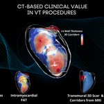
ADAS 3D helps visualize and quantify fibrosis, wall thickness, and anatomical structures to aid in pre-procedure planning and decision-making from MRI and CT
Barcelona, Spain
Joined April 2017
- Tweets491
- Following186
- Followers1.4K
- Likes377





ADAS3D Medical retweeted

Our last publication in @JACCJournals JACC EP: Ring-like pattern in NICM: agressive pattern and higher recurrence rate after RF. Understanding VT substrate with imaging. Real need for improvement. Good job of our senior fellow @RaimondoPi4757 @hospitalclinic @aportasanchez
jacc.org/doi/10.1016/j.jacep…


ADAS3D Medical retweeted
Happy to see 2 studies from @hospitalclinic in @hrs_journal same edition about#imaging in VT @ADAS3D and non-invasive mapping #ECGi to analyse substrate @corifyAF.
More to come! @janareventos @S_Vazquez_Calvo @aportasanchez

ADAS3D Medical retweeted
Redo case: ICM+VT storm. Functional #S3 map (HDGridX). DZs correlate with CMR @ADAS3D. LPs in S1, blocked in S2 visible again in S3. Critical area for VT: 99% pacemap+long S-QRS. Well done @t_rosseel ! @hospitalclinic @tolosanaviu @EduGuasch @aportasanchez


ADAS3D Medical retweeted

Chagas Megacolon in VT. Colon relationship identified using @ADAS3D software. First was a case scheduled for today and we decided not to go epi, second an old case that had an Huge megacolon that went to surgery @Dr_Santangeli @DrRoderickTung @LuigiDiBiaseMD @aalahmadmd


ADAS3D Medical retweeted

Iatrogenic substrate revisited @JACCJournals
CMR-detected post-ablation scar channels predict recurrent AF after PVI and may be an indicator of iatrogenic substrate as well as a promising ablation target. 💪@MarionaRegany @LluisMont2 @ivroca @AGEP_DGK
bit.ly/4hsuZ2Y
ADAS3D Medical retweeted

“Simplicity is the ultimate sophistication”
Left atrial atypical flutter involving posterior wall scar and right PVs.
⏳ Skin-to-skin procedure time 79 min.
🩻 Fluoroscopy <2 min.
PVI and roof line adapted to LAWT and esophagus position.
@ADAS3D @Clinica_Teknon #Epeeps

Imaging-Guided Ablation for Atypical Atrial Flutter 📡
Posterior line ablation using ADAS & CT imaging for precise lesion placement
Done with a tetrapolar catheter & SmartTouch
@JAldereteM @DrBerruezo @Davidsotoiglesi @ChiaraValeriano
#EPeeps
ADAS3D Medical retweeted

Dr. Roca-Luque @ivroca discusses the role of CMR in evaluating ablation success and RF lesion evolution at the 2024 VT Symposium. #CardioTwitter #Cardiology #EPeeps #MedTwitter
Register to watch the VT Symposium livestream now: vtsymposium.com/registration

ADAS3D Medical retweeted
Happy to share a case-in-a box of VT ablation guided by MRI + S3 protocol in @Someec1 showing how to take advange of structure information and #S3 protocol. Less tha 2 hours! @hospitalclinic @aportasanchez @barnaclinic @ADAS3D @tolosanaviu @LluisMont2 @TillAlthoff @guichard_jb


ADAS3D Medical retweeted

Our current understanding of post-MI VT substrate derives heavily from completed MI model. We used a more contemporary reperfused MI swine model w/ 90min mid-LAD occlusion and studied scar with sequential LGE-CMR, @ADAS3D , functional substrate mapping & programmed stim. 2/5

ADAS3D Medical retweeted

Brilliant work shared by @ivroca excited for what's next.
#EnSite S3 DeEP Mapping techniques & preprocedural imaging with @ADAS3D.
VT In-Depth by @JRWinterfield Dr. Wendy Tzou, et al.
@AbbottCardio

ADAS3D Medical retweeted

With an expanded industry-led, hands-on skills program planned for #HRS2024, we're seeking the best that industry has to offer! Submit your session proposals by Nov 17 for the chance to showcase how YOUR products uniquely fill #EPeeps' needs.
LEARN MORE > bit.ly/3s2G4Us

ADAS3D Medical retweeted

🎯 Highly Individualized PVI approach guided by left atrial wall thickness
#EUROPACE_FRESH_OFF_THE_PRESS
⚡️see data from Spain now available in #Europace
👉doi.org/10.1093/europace/eua…
@GiulioConte9 @Dominik_Linz @marcovitoloMD @AndyZhangMD @FraSantoroMD

ADAS3D Medical retweeted

Computed tomography-guided personalized ablation for persistent atrial fibrillation at @Clinica_Teknon.
By-LAWT protocol by @DrBerruezo @diegopenela1 @Davidsotoiglesi @julio93383971 @DrCherylTeresEP @pietro_francia @AndreaSaglietto @JAldereteM
academic.oup.com/europace/ad…

ADAS3D Medical retweeted

Great to have @ivroca and @aportasanchez from @hospitalclinic Barcelona in our EP session today discussing OT Near field mapping. #hospitalStaCruz @cavaco_diogo @DanielM891

ADAS3D Medical retweeted
More and more often we perform highly invasive redos only to find all 4 PVs isolated…
…LGE-MRI can rule out PV reconnection non-invasively. Clear potential to guide patient selection & ablation strategy for redos!
@LluisMont2 @nmarrouche @Phiso_de
bit.ly/3BR9sPw


ADAS3D Medical retweeted
Testing efficacy of catheter ablation by non-invasive lesion assessment!
bit.ly/3INZyQI
@Phiso_de @chris_sohns @EuropaceEiC @ESC_Journals @AGEP_DGK @YoungDgk @LluisMont2 @ivroca @ADAS3D @AG21_DGK @AMCliment @Dominik_Linz @DavidDuncker @escardio @aportasanchez

ADAS3D Medical retweeted

More evidence for CT-derived Left Atrial Wall Thickness maps for #AFib ablation. @ADAS3D #EPeeps #Cardiology #CardioTwitter #EPBot

🚨Latest #FreeRead Article in
@JICE_EP
Reproducibility Analysis of the CT Angiography–Derived LA Wall Thickness Maps
A Valles-Colomer, BR Forcada, @Davidsotoiglesi, @DrCherylTeresEP @diegopenela1 @DrBerruezo & RM Figueras i Ventura
📖🧐: rdcu.be/c55aC
#EPeeps


ADAS3D Medical retweeted

Our first @ADAS3D case. ICM, lateral scar. Great first impressions. Showed deep endo location of main channel. Outstanding support from @RosaMFiV. @diananguyenn @_nataliecohen95 @Ghassanj89 figured out integration with CARTO, great team work.