

PostDoc Institut of plant science
- Tweets115
- Following201
- Followers159
- Likes511






ALT Dr Lucie Riglet developed a quantitative imaging pipeline and deciphered the mechanisms specifying the distinct bullseye regions in developing petals of Hibiscus trionum combining imaging, genetics, computational modelling and bumblebee behaviour assays. (A) Hibiscus trionum petals with a boundary at the one-third position separating the purple proximal region and white distal domain. (B) At early developmental stages, when the petal is still greenish, lacking of any differentiation sign, a pre-pattern is already specifying the boundary region of the petal at maturity. (C) The Moyroud team identified the mechanisms behind pattern variations, using Hibiscus richardsonii, a sister species. (D) To investigate if bullseye proportion was able to be detected they tested bumblebees with artificial flowers mimicking H. trionum (medium) and H. richardsonii (small) bullseye sizes, and found that bumblebees preferred the medium-sized bullseye. Image credits: Lucie Riglet.


ALT Schematic displaying stomatal development in wild type (WT). Stomatal development starts with guard mother cell (GMC; blue) formation. BdMUTE is expressed in the GMC (light blue nucleus) and moves into the lateral cells (light yellow nucleus) to establish subsidiary mother cells (SMCs). SMCs then polarize (yellow gradient) and divide asymmetrically to form lateral SCs (yellow). Finally, the GMC divides symmetrically to generate two GCs (light green), which form a pore and mature into the characteristic dumbbell shape (dark green). Open questions regarding BdMUTE’s roles and mechanisms are indicated.


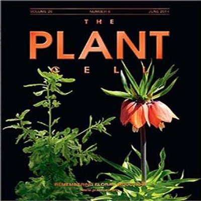


ALT Mathilde Chopy (she/her) is the First Author of Cell layer-specific expression of the homeotic MADS-box transcription factor PhDEF contributes to modular petal morphogenesis in petunia published 06 October 2023. She is an Engineer in molecular pathology at Institut Bergonie (France, Bordeaux). In 2023, Chopy did a Post-doc at Bern University. She received her PhD in 2019, studying Flower development at tge Laboratory of Plant Development and Reproduction, ENS Lyon. Chopy also holds a MSc in Biology and Biotechnology of Plants from Bordeaux University (2016). Research interests include flower development, and personal interests include ballet and salsa.

ALT Title: ROLE OF CELL LAYERS IN PETAL MORPHOGENESIS. On the cover: Petunia petals form a limb and tube through congenital fusion, a complex morphology that coevolved with pollinators. Chopy et al. (pp. 324–345) analyzed mutants (periclinal chimeras) in which the expression of the homeotic petal identity factor PhDEF is limited to either the epidermis or to the inner cell layers, with dramatically different outcome on limb and tube development. The image shows a chimeric petunia flower in which PhDEF is expressed normally in the upper petal, while restricted to the innercell layers in the four remaining petals. Image credit: Michiel Vandenbussche.


ALT Image Source: Supplemental Data. Chopy M. et al. (2023). Role of cell layers in petal development. Plant Cell. https://academic.oup.com/plcell/advance-article/doi/10.1093/plcell/koad258/7296041 Supplemental Figure S1. Additional pictures of star and wico flowers (Supports Figure 1). Star (A-D) and wico (E-I) flowers from independent branches, viewed from the side (left) and from the top (right). Sepals have been occasionally removed for clarity. Scale bars: 1 cm.



ALT One gene, two mutations, contrasting phenotypes. Model to illustrate EOB2 multi-functionality. The knockout (KO) lines and LofTAD lines do not present the same floral phenotype, while both lines are unable to activate target genes (represented by blunt-ended arrows). This is the proof that EOB2 is involved in other functions.


