

High-Fidelity Simulators for Neurosurgeons and Interventional Neuro Radiologists: Patient-Specific Treatment Planning and Strategy for Complex Cases
Dallas, Texas, USA
Joined April 2020
- Tweets788
- Following2K
- Followers1.5K
- Likes22.4K






78 years old. A minor fall at home, a burr-hole evacuation, and three months later the repeat CT shows the subdural is back, with midline shift again. The neomembrane has re-bled, fed by fragile micro-vessels off the middle meningeal artery. This time the team wants to treat the source too.
In the angio suite, the microcatheter makes the turn from the internal maxillary artery into the MMA and heads up toward the foramen spinosum. From there, every injection is a judgment call. Push the liquid embolic too hard and it refluxes where you don’t want it. Hold back too much and the dural bed keeps feeding the membrane.
Dural anatomy varies a lot from one patient to the next, and liquid embolics need a stable microcatheter and a patient hand. Both are far easier to learn on the bench than under live fluoro.
That’s what we’re bringing to the @svinsociety (Society of Vascular and Interventional Neurology) 2026 Annual Meeting in Las Vegas: our MMA embolization models for advanced hands-on training. They’re built from patient imaging and fully transparent, so you can watch exactly where the embolic goes. And they work with the agents physicians use clinically: Onyx®, Squid®, PHIL, n-BCA and coils.
→ Track your microcatheter through tight dural branches
→ Practice slow, controlled embolic delivery
→ Find the point where reflux starts, before you find it in a patient
📅 18–20 November 2026
📍 SVIN26 Annual Meeting, Las Vegas, Nevada
Heading to Vegas? Bring the microcatheter and embolic you actually use and run them through the model with us.
And we’re curious: for a recurrent cSDH, what drives your choice between EVOH, PHIL and n-BCA?
#SVIN26 #MMAEmbolization #ChronicSubduralHematoma #Neurointervention #InterventionalNeuroradiology #Embolization #MedicalSimulation #ProceduralTraining

137
The microcatheter tip is sitting at the apex of the carotid siphon. Below it, a bovine arch that fought you the whole way up. On fluoro, every millimeter of wire feels huge and the clock is running. It’s an LVO, and you don’t get a practice run in a living patient.
Anyone who has done enough thrombectomies knows that moment. The resistance in your hands. The small corrections between the aspiration catheter and the stent retriever. That’s often the difference between a clean first pass and a long fight. It’s exactly the moment we want physicians to rehearse before they walk into the angio suite.
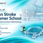
This week we’re heading to Thiruvananthapuram for the Asian Stroke Summer School 2026, supported by the Sree Chitra Alumni Association of Neurology (SCAAN) and organized by the Comprehensive Stroke Care Program at SCTIMST Clinical Engineering and the Stroke Center at Universitätsklinik für Orthopädische Chirurgie und Traumatologie Inselspital Bern, University of Bern, under course directors Dr. PN Sylaja, Dr. ER Jayadevan, Dr Urs Fischer and Dr. Jan Gralla alongside faculty including Dr. Blaise Baxter and Dr. Johannes Kaesmacher. It’s endorsed by ESMINT Society, INDIAN STROKE ASSOCIATION, European Stroke Organisation, Society of Vascular and Interventional Neurology (SVIN), World Stroke Organization and WFITN, and we’re proud to support the hands-on sessions.
Participants will work on our flow-perfused neurovascular models to:
→ Run the full EVT workflow, step by step, under realistic flow and pressure
→ Work through tortuous arches and difficult access before meeting them in a high-risk case
→ Compare device handling and retrieval techniques, and repeat a pass as often as they need
📅 12–15 November 2026
📍 Thiruvananthapuram, Kerala, India
Coming to Kerala? Which access problem would you most like to rehearse on the model? Tell us in the comments, or DM us to meet the team on-site.
#AsianStrokeSummerSchool #Neurointervention #EndovascularThrombectomy #MechanicalThrombectomy #StrokeCare #InterventionalNeuroradiology #MedicalSimulation #SCTIMST #Inselspital #SurgeonsLab

1
120
For years, the venous side was the part of the run most of us scrolled past.
That's changing fast. This year the FDA approved the first stent designed specifically for the cerebral venous sinuses. Dedicated venous stents for pulsatile tinnitus are in trials. Endovascular CSF shunts are being placed through the inferior petrosal sinus. And randomized trials like ESCORT and REVIVE-CVST are testing whether thrombectomy should become standard care for severe Cerebral Venous Sinus Thrombosis (CVST).
Every one of those devices has to work in the same awkward place. Up from the femoral or jugular vein, through the jugular bulb, round the sigmoid bend, into a transverse or sagittal sinus that may be packed with clot. Thin walls, variable anatomy, and a lot less hands-on training than the arterial side gets.
So that's where we started with our CVST model.
We took DICOM data from multiple centers, plus anatomical sketches of the detail imaging alone doesn't capture well, and printed it first in clear resin. That let us check geometry and catheter paths before committing. Then we moved it into flexible silicone on a flow loop, with the dural venous sinuses and the Circle of Willis in the same circuit.
On the bench, teams can:
→ track microcatheters and guides through tortuous venous pathways under fluoroscopy
→ rehearse thrombectomy and recanalization techniques with physiological flow
→ see what venous outflow restriction does to pressure across the sinus
→ run the same case again and again until it's routine
We built it for fellowship programs, for device teams working on venous stents, thrombectomy tools and transvenous implants, and for centers that want a patient-specific version before a difficult case.
If you work on the venous side, what's been hardest to test or teach so far? Tell me below, or message me if you need a custom venous model.
#InterventionalNeuroradiology #Neurovascular #CVST #IIH #VenousSinusStenting #PulsatileTinnitus #MedicalSimulation #MedicalDeviceDevelopment #MedTech #SurgeonsLab @serenityiih @CereVasc @SNISinfo @svinsociety @esmintsociety
3
228
A PE case rarely gives you time to think twice. Clot burden, a strained right heart, a wire that won't go where you want it. Decisions happen fast, and the anatomy doesn't forgive hesitation.
That's the pressure we want people to feel at AHA Scientific Sessions 2026.
SurgeonsLab is bringing its Pulmonary Embolism Simulator to the Simulation Zone (#120) for all three days. It's a high-fidelity heart and pulmonary vasculature model running on a physiological flow loop, so you work through the procedure the way it actually unfolds.
At the bench you can:
→ Navigate catheters from the right heart into the pulmonary arteries
→ See how flow hemodynamics change device behavior
→ Run thrombus retrieval protocols in realistic anatomy
It's built for interventional physicians, clinical educators and device teams who want to test technique or a device on patient-specific flow models before it matters most.
Need different anatomy or a specific test setup? We also take on custom models and training programs.
AHA Scientific Sessions 2026
Simulation Zone, #120
McCormick Place, Chicago, IL
November 7–9, 2026
Heading to Chicago? Drop a comment or send us a message to book a dedicated hands-on session. Bring your device if you want to put it through a run.
#AHA26 #PulmonaryEmbolism #InterventionalCardiology #InterventionalRadiology #Endovascular #MedicalSimulation #MedTech #MedicalDevices #ProceduralTraining #SurgeonsLab
2
172
In January, a neurosurgery team from Children's Cancer Hospital - Egypt 57357 visited us in Bern with one wish: a place in Cairo where physicians could train all year.
In September, that lab opened.
One room, four stations:
- Brain tumor surgery and aneurysm clipping under the microscope
- Microvascular anastomosis on NeuraPass models, with vessels from 0.4 to 1.5 mm
- Catheter navigation and coiling on patient-specific endovascular flow models
- 40 neurovascular cases in CathTrain VR, endovascular virtual simulator
What stayed with us most: the faculty didn't only want to use the lab. They asked to learn how to set it up, reset it and run it themselves. That is what keeps a training center open all year.
We've written up the whole journey, from the first conversation in Switzerland to installation week in Egypt, with seven practical lessons for anyone planning a simulation lab.
Read the story: surgeonslab.com/blogs/cche-5…
Thank you to the faculty and teams at CCHE 57357 for the warm welcome in Cairo.
#Neurosurgery #SurgicalSimulation #MedicalEducation #Neurointervention #Egypt

1
122
Clip’s on, aneurysm still filling on ICG. Your next move?
Teaching points from a hands-on clipping course at King’s College London, taught on our patient-specific MCA aneurysm model.
(Brain colours softened for social media. The real model is more lifelike)
#Neurosurgery #AneurysmClipping
A bag of saline on an IV pole gives you flow. It doesn't give you a pulse.
You notice the difference as soon as a device goes in. A microcatheter tip, a coil or an aspiration catheter behaves differently in a vessel that moves 70 times a minute than in one sitting under steady pressure. If you're teaching embolization, the way the embolic travels downstream depends on that pulse too.
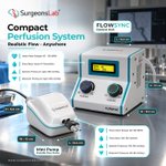
The pump also has to fit the job. A full vascular loop on a test bench needs more than a single model on a workshop table, so we built two compact pulsatile units.
FLOWSYNC Control Unit - For full circuits and models with higher flow needs
- Dimensions: 16.5 x 18.5 x 16 cm
- Pulse rate from 40 to 120 BPM
- Flow up to 0.7 L/min
- Systolic up to 160 mmHg, diastolic up to 100 mmHg
Mini Pulsatile Pump - For smaller models, focused sessions and portable workshops
- Dimensions: 8.5 x 12 x 6.5 cm
- Pulse rate from 60 to 100 BPM
- Flow rate from 160 to 190 mL/min
- Systolic 110 to 130 mmHg, diastolic 60 to 80 mmHg
On both, you set pulse rate, pressure and flow yourself.
On the FLOWSYNC you can run a model at 45 BPM, then take the same anatomy up to 160 systolic and watch what changes. They sit alongside the continuous-flow setups in our perfusion range, for models where steady flow is all you need.
Built for:
→ Interventional radiologists and course directors running embolization and endovascular workshops
→ Device engineers comparing designs under the same, repeatable conditions
→ Field and clinical education teams taking a working demo to a hospital or congress
Planning a workshop, a device demo or a new bench study? Tell us the model and the flow you need, and we'll help you pick the right setup. Visit: surgeonslab.com/mini-perfusi…
What's the one thing your current flow setup can't do that you wish it could?
Setting up a vascular model, a test bench or your next workshop? Send us the application, the model size and the flow and pressure you need, and we'll suggest a configuration that fits.
Email us at [email protected] or Visit surgeonslab.com/contactus
#InterventionalRadiology #Endovascular #Embolization #Hemodynamics #MedicalDeviceTesting #SurgicalSimulation #MedicalSimulation #SurgeonsLab

127
Heading to MD&M Midwest in Minneapolis? Come find us at Booth #2946.
If you're developing a device for spine, Vascular, peripheral or oncology procedures, you already know how far a benchtop fixture is from a real patient. That gap is exactly where we work.
At SurgeonsLab, we build simulation models that look, feel and behave like real anatomy. That means tissue with realistic feel, vessels with physiological flow, and anatomy that responds the way your device will meet it in the clinic.
Device teams work with us across the whole product lifecycle:
• R&D and early design iteration
• Bench testing and design verification
• Human factors and usability studies
• Physician training and procedural education
• Sales enablement and live demos that actually show your device in action
Whether you're working on a Class I instrument or a Class III implant, we can build around your device, your procedure and your target anatomy, including patient-specific models from imaging data.
At the booth you'll get hands-on time with:
Surgical Spine Model
Vascular & Tumor Model
Reusable Embolic Demonstrator (RED)
CathTrain VR endovascular simulator
Endovascular flow models with Circle of Willis aneurysms
What we'd most like to hear about is the procedure you're struggling to test, teach or demo. Bring it to us and let's talk about building it.
Flying in from the East Coast or elsewhere? Send me a message and we'll set a time to meet so you're not hunting for us on the show floor.
Booth #2946, Minneapolis Convention Center
October 28–29, 2026
#MDMMidwest #MedTech #MedicalDevices #MedicalSimulation #DeviceDevelopment #InterventionalRadiology #Endovascular #Neurovascular #SpineSurgery #HumanFactors #MedicalAlley #SurgeonsLab

1
114
Getting the embolic in is the easy part. The real question is where it goes next.
In MMA embolization, "how distal is distal enough?" has become one of the most debated questions in the field. Go deeper and you may cover more of the target territory. Push too hard and you risk reflux back along the catheter, or flow into places you never meant to reach.
The tricky part is that a lot of this happens in vessels smaller than a millimeter, where it's hard to see what's actually going on.
So we filmed it. This video is live fluoroscopy of a liquid embolic moving through our MMA model, with branches in the sub-millimeter range. You can watch two things happen in real time:
1. How far the embolic penetrates into those small distal branches
2. When reflux starts, and how far it travels back toward the catheter tip
Because the anatomy is the same every run, you can change one thing (injection speed, catheter position, the agent itself) and see exactly what that change does.
For physicians, that means practicing the feel of a controlled injection before it matters. For device teams, it means comparing how an agent or catheter behaves in a consistent, repeatable anatomy.
Watch it, and tell us: what would you test first?
#MMAembolization #Neurointervention #Embolization #Endovascular #InterventionalNeuroradiology #MedicalSimulation #MedTech #SurgeonsLab
1
145
Evaluating embolic penetration and reflux dynamics requires bridging direct bench-top observation with real-world clinical imaging.
The Reusable Embolic Demonstrator (RED) provides a single, flow-enabled platform supporting three distinct visualization modalities:
Optical Camera: Real-time, transparent tracking of vessel filling, reflux patterns, and occlusion mechanics.
Fluoroscopic: Seamless integration for embolic tracking and workflow evaluation in an angio suite
CT road mapping: High-resolution cross-sectional computed tomography imaging for detailed 3D volumetric assessment.
Pairing quick-swap pathology modules with multi-modal compatibility gives R&D and clinical teams a cost-controlled setup to validate embolic behavior from benchtop to angio suite.
Know more about the Reusable Embolic Demonstrator, visit surgeonslab.com/reusable-emb…
You can also drop us an email at [email protected]
#Endovascular #Neuroradiology #LiquidEmbolics #SurgeonsLab #MedicalDeviceTesting #Neurointerventions

79
Evaluating embolic penetration and reflux dynamics requires bridging direct bench-top observation with real-world clinical imaging.
The Reusable Embolic Demonstrator (RED) provides a single, flow-enabled platform supporting three distinct visualization modalities:
• Optical Camera: Real-time, transparent tracking of vessel filling, reflux patterns, and occlusion mechanics.
• Fluoroscopic: Seamless integration for embolic tracking and workflow evaluation in an angio suite
• CT road mapping: High-resolution cross-sectional computed tomography imaging for detailed 3D volumetric assessment.
Pairing quick-swap pathology modules with multi-modal compatibility gives R&D and clinical teams a cost-controlled setup to validate embolic behavior from benchtop to angio suite.
#Endovascular #Neuroradiology #LiquidEmbolics #SurgeonsLab #MedicalDeviceTesting #Neurointerventions

101
Advancing neurovascular training standards across the Middle East requires simulation infrastructure capable of replicating authentic anatomical friction, microscopic depth, and tactile feedback.
SurgeonsLab is pleased to announce the successful installation and operational setup of our SurgTrain surgical platform, endovascular aneurysm models and CathTrain VR at Children’s Cancer Hospital Egypt 57357 (CCHE 57357)—a premier regional center for pediatric and neuro-oncology care.
The installed suite provides CCHE 57357 with a comprehensive physical simulation environment:
SurgTrain Surgical Console: Designed for high-fidelity microsurgical clipping practice under microscope visualization, replicating realistic tissue resistance and spatial constraints.
Benchtop Endovascular Aneurysm Models: Configured for microcatheter manipulation, coil deployment, and navigating complex vessel tortuosity.
Neura-Bubble & Modular Units: Specialized setups engineered for targeted anatomical access and procedural refinement.
As healthcare institutions across Saudi Arabia and the broader MENA region continue to elevate clinical simulation infrastructure, establishing patient-specific, reusable dry-run environments is essential for building procedural confidence without intraoperative risk.
For neurosurgery and interventional radiology leads in the region: How is your institution incorporating physical simulation suites into resident training and complex case planning?
To explore bringing SurgeonsLab neuro-simulation platforms to your hospital or training center in the Middle East, contact our team at [email protected] or visit surgeonslab.com/contactus
#Neurosurgery #InterventionalRadiology #Neurovascular #SurgicalSimulation #MedTech #SaudiHealthcare #MENAHealthcare #CCHE57357 #MiddleEastMedTech #SurgeonsLab #SurgTrain #CathTrainVR #ClinicalTraining
2
155
Absolutely. The value of a realistic bench model is not just in reproducing anatomy, but in giving clinicians the opportunity to work through the technical challenges before they encounter them in the angio suite.
For us, that means realistic anatomy, repeatable device interaction, and measurable bench performance—so teams can train, evaluate, and refine technique in a controlled environment.
Appreciate you highlighting this. We’re continuing to build around that principle at SurgeonsLab.
Explore our endovascular simulation flow models, visit:
surgeonslab.com/endovascular…

8
Glue or Onyx? Coils or particles? How distal is distal enough?
MMA embolization is everywhere in neurointervention right now, and good operators still disagree on how to do it. What nobody disputes is that it's a small, tortuous vessel with very little room for error.
So we built a model for it. True-to-scale MMA trunk and branches, validated with DMSO and nBCA, angio-ready, in 2D and 3D, single-use or reusable, with an optional closed-loop perfusion setup. Navigate, coil, glue, inject particles, then do it again.
For fellows, for experienced operators trying something new, and for device teams who need consistent anatomy.
Want one for your course or lab? Send us a message.
#MMAembolization #Neurointervention #Neurosurgery #MedicalSimulation #MedTech #SurgeonsLab

1
197
Understanding vascular anatomy on 2D imaging is straightforward. Navigating that anatomy under a surgical microscope is where procedural complexity begins.
During the 9th Walter E. Dandy India Neurosurgical Educational Course in Siliguri, the trainees got hands-on experience with complex aneurysm geometries and navigating deep surgical corridors. While pre-operative planning clearly outlined vessel connections, translating that plan to microscopic execution introduced real-world physical constraints: shifted working angles, restricted depth, vessel compliance, and precise clip orientation.
This gap between imaging and physical manipulation is why hands-on simulation on our SurgTrain Surgical Simulator is necessary for neurosurgeons and residents to practice patient-specific vessel tortuosity, testing force application, micro-dissection angles, and clip trajectories in a controlled environment.
True surgical readiness builds from refining and repeating techniques on the SurgTrain platform until execution is natural.
Our sincere thanks to the Walter E. Dandy India Neurosurgical Society, distinguished faculty, and participating trainees for an outstanding educational session.
To discuss patient-specific simulator models for your institution, connect with us at [email protected].
You can also connect with us at surgeonslab.com/contactus
#SurgeonsLab #WalterEDandy #Neurosurgery #SurgicalSimulation #NeurosurgicalEducation #Microsurgery #SurgicalTraining #MedicalSimulation #Neurovascular #ACOM #ICA #MCA #TumourResection #Neuro #Surgery Walter E. Dandy Neurosurgical Students Club India
137
September is Brain Aneurysm Awareness Month.
A brain aneurysm is a weakened area in a cerebral artery wall that can create a complex clinical and procedural challenge.
1 in 50 individual have an unruptured cerebral aneurysm. When the aneurysm ruptures, the clinical impact is immediate—a critical stroke event carrying a 50% mortality rate.
Treatment may involve coiling, stent-assisted coiling, flow diversion, or surgical clipping, depending on the aneurysm and the clinical situation.
For neurovascular teams, whether deploying endovascular techniques like coiling, stent-assisted coiling, and flow diversion, or executing microsurgical clipping, navigating complex vessel tortuosity and delicate wall geometries leaves zero margin for error. Operative success relies on device behavior, precise positioning, and thorough anatomical familiarity before making the first access site.
This Brain Aneurysm Awareness Month, the focus must extend beyond detection to procedural mastery.
Rehearsing complex anatomy and device–anatomy interaction in a controlled environment can support technical training and procedural mastery.
At SurgeonsLab, we develop neuro-simulators designed to help teams rehearse, refine challenging neurovascular procedures and validate neurovascular devices faster.
This month is an opportunity to raise awareness, support education, and keep advancing preparation for complex cases.
Rehearse the anatomy. Refine the technique. Be ready for the procedure.
#BrainAneurysmAwarenessMonth #BrainAneurysm #Neurovascular #Neurointervention #MedicalSimulation #EndovascularTraining #SurgeonsLab #MedTech

1
102
5,000+ Followers.
A small number on a screen, but a meaningful milestone for the SurgeonsLab community.
Thank you to the surgeons, interventionalists, educators, biomedical engineers, device professionals, researchers, and MedTech teams who follow our work, challenge our thinking, and engage with what we share.
We started SurgeonsLab around a simple idea: realistic simulation should bring anatomy, devices, and procedural technique closer together.
That means more than replicating anatomy.
It means creating models where complex neurovascular anatomy, tortuosity, bifurcations, access challenges, and device–anatomy interaction can be explored in a controlled and repeatable environment.
Whether it is evaluating microcatheter navigation through acute bifurcations, studying embolic delivery in MMA models, or refining access techniques, the objective remains the same:
Understand the procedural challenge. Test it repeatedly. Generate meaningful evidence before it reaches the patient.
Over the past few years, the conversations with this community have helped shape the questions we ask and the problems we work to solve.
5,000 is not the destination. It is a reminder to keep building, testing, learning, and sharing.
Thank you for being part of the journey.
— SurgeonsLab
#SurgeonsLab #MedicalSimulation #MedTech #SurgicalTraining #EndovascularTraining

78
Spot-on observation @dtruscello215
Navigating 2nd and 3rd order intrahepatic ducts remains a major ERCP bottleneck—standard cannulas often lack the torque and stability for acute, tight branches.
Translating microcatheter mechanics into interventional GI brings three key advantages:
1:1 Torque Control: Selective entry into tight side-branches without wire buckling.
High Trackability: Superior column strength to cross fibrotic strictures.
Low Profile: Reduced mechanical trauma in sub-centimeter channels.
Bench-validating these navigation dynamics on anatomical models is the critical first step toward proving cross-disciplinary feasibility.
1
35
We’ve all been there @DrPsylocibo . The hydrophilic wire helps, but that 90° turn still makes you earn it.
1
16