


faei retweeted

Just submitting a paper with this group and have run the samples literally from this study on our platforms thanks to a connection from @ryan_cxm 😎
Autoantibodies are partially reset, level of that correlates with outcome.
faei retweeted

We’ve been blown away by the response to my interview with @BBCSport & @KatieFalkingham , thank you so much to everyone who has taken the time to read and engage with it ❤️ millions of views across multiple platforms really is immense.
We have since written to @andyburnham , calling on the @UKLabour government to fund research into this horrific illness. I know there’s so much more we could say, but with limited capacity, we’ve tried to focus on the lack of funding, resources in the NHS and how much it’s actually costing the government.
As you can see on the slides, it has been drastically underfunded compared to other illnesses and it is currently costing the UK Government far more than it would cost to fund research. ME is most often a post-viral illness. Therefore, since the start of the Covid pandemic, we’ve seen cases increase drastically and they will continue to increase. We need funding into research urgently. With estimates of people with ME being around 1.35 million, this is a lot of lives destroyed.
I’ve had thousands of messages on multiple platforms since sharing my story so please bear with me ❤️
Thanks to @ThereForME_UK and @wecrunchme for the stats.


faei retweeted

‼️Remember our hypothesis of autoimmune hypophysitis in ME/CFS and Long COVID, published in 2024?
We proposed that infection-driven immune hyperactivation, genetic susceptibility and loss of immune tolerance could contribute to pituitary dysfunction, ACTH deficiency and hypocortisolism.
Now, the GEM genetic study reports the pituitary gland as the most enriched tissue.
This does not confirm autoimmune hypophysitis, but it strengthens the rationale for investigating a possible neuroimmune-endocrine mechanism in a subgroup of patients.
doi.org/10.3389/fimmu.2024.1…


4) One of the more interesting findings is is that the pituitary (green bar below) now stood out clearly in the tissue analysis. This may point to an important role for hormones in the disease.
See our recent article on prolactin response in ME/CFS here:
mecfsscience.org/increased-p…

faei retweeted

Interesting: older findings in #MECFS show a markedly increased prolactin response to buspirone:
mecfsscience.org/increased-p…
A new genetic study now points to the pituitary in the tissue analysis:
teapawt.net/mecfsskeptic/status/21…
A good lead for further studies!


faei retweeted
⚠️I think there are several problems with that reasoning.
First, there is a fundamental mistake in assuming that because more than 90% of adults are infected with EBV, everyone is equally capable of controlling the virus.
This is precisely where one of the similarities we have been discussing for years comes in.
A subset of people develop infectious mononucleosis, characterized by a massive expansion of CD8+ T cells, intense immune activation, and symptoms such as fatigue that can persist for months.
Moreover, some patients subsequently develop ME/CFS, a disease for which we have long proposed a possible autoimmune origin, associated with an immunogenetic susceptibility shared with other autoimmune diseases.
In our 2025 publication, we proposed that certain ancestral HLA class II haplotypes could favor abnormal immune responses to persistent antigens, contributing to immune hyperreactivity, functional exhaustion, and loss of immunological tolerance.
So what happens when the immune system continues responding to viral antigens that it cannot adequately control?
In genetically susceptible individuals, this stimulation could sustain a disproportionate immune response that, over time, contributes to T-cell dysfunction and the development of autoimmunity.
Sound familiar?
This is precisely one of the mechanisms we proposed for long COVID back in 2023.
Although the pathogens are different, our hypothesis is that certain individuals may share an immunogenetic susceptibility that predisposes them to develop pathological immune responses to persistent infectious stimuli.
The difference is that EBV has evolved specific mechanisms to establish lifelong latency, whereas SARS-CoV-2 has no known equivalent latency mechanism.
In the case of SARS-CoV-2, some people may completely clear the infection, while others may retain very small amounts of virus or viral material in certain tissues, such as mucosal tissues, without necessarily having ongoing active replication.
And this is where I think one of the key issues lies.
Even very small amounts of persistent viral antigens could contribute to sustaining a disproportionate immune response in individuals with an immunogenetic susceptibility shared with other post-infectious autoimmune diseases.
In our model, certain ancestral HLA class II haplotypes could favor this hyperreactivity to antigenic stimulation, contributing to prolonged immune activation, T-cell dysfunction, and loss of tolerance.
This could help explain why some people can keep potential viral reservoirs under control without developing disease, while others may experience a pathological immune response even in the presence of a relatively low antigenic burden.
Second, the fact that antivirals cannot completely eliminate a persistent infection does not mean they are useless.
An antiviral can reduce viral replication and, when active viral production is occurring, decrease the amount of viral antigen sustaining the immune response.
Of course, if only non-replicating viral material persists, conventional antivirals may have limited usefulness. But we cannot assume that every patient has the same type of persistence or that all reservoirs have the same biological activity.
The examples of multiple sclerosis and lupus are particularly interesting.
There is very strong evidence supporting a causal relationship between EBV infection and multiple sclerosis, while substantial evidence also links EBV to the pathogenesis of lupus. Associations between EBV activity and disease flares are also being investigated.
If viral reactivations contribute to sustaining or worsening certain inflammatory processes, why wouldn’t it be worth trying to control them?
That does not mean it has already been demonstrated that currently available antivirals prevent multiple sclerosis or lupus flares. We still need clinical trials to establish that.
1/3👇🏻

Viral persistence in COVID is different from that of viruses like EBV & HIV. Those viruses persist for 99%+ of people.
Covid only seems to persist for a subset from all available research. This means that the solution to COVID persistence can be different.
The persistence most likely will be caused by a defect in the immune system, rather than a specific nature of the virus. This means that the solution may need to be restoring the immune system (potentially stuff like checkpoint inhibitors etc - but research needed), rather than antivirals.
If the virus itself is not replicating, then antivirals will not do much. The persistence would be caused by a lack of clearance capability. That is what we need to fix.
At least until we can make drugs that can actually remove the virus directly on a systemic level. I imagine AI will help with this very soon. But it will still have to go through clinical trials, which is the long part.
faei retweeted
🔴 What If Different Infections Could Trigger the Same Immune Dysfunction? New Evidence on Long COVID, EBV, and T Cell Exhaustion
Why do some people never fully recover from an infection? Why can viruses as different as Epstein–Barr virus (EBV) and SARS-CoV-2 trigger prolonged illnesses with similar clinical and immunological features?
And, perhaps more importantly, what happens when the immune system remains activated for months or even years but fails to eliminate the stimulus driving that response?
These questions have guided much of our research over the years on myalgic encephalomyelitis/chronic fatigue syndrome (ME/CFS), long COVID, and autoimmune diseases.
Since 2021, we have been developing an immunopathological model linking viral persistence, immune evasion mechanisms, T cell hyperactivation, and subsequent immune dysfunction to the development of certain chronic post-infectious illnesses.
Now, a new study by Painter and colleagues, published on October 16, 2026, provides experimental findings that align with several mechanisms we proposed in our earlier work. And there is one particularly interesting connection worth revisiting.
In our first paper, published in Frontiers in Immunology in 2021, we proposed that certain patients with ME/CFS might have difficulties controlling EBV-infected cells that remain in a latent state, particularly in the presence of specific genetic susceptibility factors related to HLA class II.
EBV can persist in the body for life, primarily within memory B cells. Under normal circumstances, the immune system keeps these infected cells under control. However, the virus has evolved several immune evasion mechanisms that allow it to interfere with immune recognition and clearance.
Our hypothesis suggested that insufficient immune surveillance could favor the persistence of infected cells and contribute to chronic immune abnormalities.
In 2022, we explored this model further, focusing on the potential role of cytotoxic CD4+ T cells in the surveillance of latently EBV-infected cells, particularly those expressing antigens such as EBNA-1.
This distinction matters because EBV control does not depend exclusively on CD8+ T cells. CD4+ T cell responses also play important roles in recognizing infected cells, and alterations in these populations could facilitate viral immune evasion.
The central idea was that the problem might not be limited to the initial infection itself, but also involve the consequences of inadequate immune surveillance against a pathogen capable of persisting and reactivating.
Our publications from 2021 and 2022:
doi.org/10.3389/fimmu.2021.6…
doi.org/10.3390/pathogens110…
However, it was in September 2023 that we explicitly extended this model to long COVID.
In a paper published with Bruno Paiva and Aintzane Zabaleta in the Journal of Translational Medicine, we proposed that an insufficient antiviral CD4+ T cell response, potentially influenced by certain HLA class II haplotypes, could favor SARS-CoV-2 persistence in tissue reservoirs, leading to prolonged antigenic stimulation.
But we also considered what this sustained stimulation could mean for T cell function.
We proposed that chronic exposure to viral antigens could impair the generation of memory T cells, promote differentiation toward short-lived effector populations, and progressively lead to states of functional T cell exhaustion.
In other words, an immune response that remains activated but gradually loses part of its ability to control the very stimulus that keeps it going.
And we went one step further. We suggested that this deterioration in immune function could compromise surveillance against other persistent viruses, particularly EBV, potentially facilitating its secondary reactivation.
Under this model, an initial SARS-CoV-2 infection could create an environment of prolonged immune stimulation while simultaneously impairing the control of another virus that is normally kept under surveillance.
This is not an interpretation we are making retrospectively in light of the new findings. It is a hypothesis we explicitly developed in our 2023 paper.
doi.org/10.1186/s12967-023-0…
Three years later, Painter and colleagues have analyzed virus-specific T cell responses in more than 500 blood samples collected from over 300 participants, including individuals with long COVID and recovered controls.
Using spectral flow cytometry, peptide-HLA tetramers, and single-cell sequencing, they identified T cells targeting different viruses and examined their functional characteristics.
Their approach is particularly interesting because they did not simply assess generalized immune activation. Instead, they were able to identify which T cell populations were activated and which viral antigens those cells recognized.
They found that 40% of samples from individuals with long COVID showed elevated activation of SARS-CoV-2 spike-specific CD8+ T cells, compared with 16% of samples from recovered controls.
Importantly, this activation could persist for months and was accompanied by specific functional alterations.
Spike-specific CD8+ T cells displayed transcriptional signatures consistent with persistent TCR stimulation, increased effector and cytotoxic differentiation, reduced memory-associated programs, and expression of exhaustion-related genes such as TOX and LAG3.
These cells also showed a reduced ability to produce IFN-γ and TNF and impaired proliferative responses following antigen stimulation.
In other words, they continued to display signs of activation, but part of their functional capacity had become compromised.
A strong immune response is not necessarily an effective one. When antigenic stimulation persists, certain T cell populations may remain activated while developing functional abnormalities that limit their ability to control the stimulus driving their activation.
This combination of prolonged antigenic stimulation, loss of memory-associated features, and T cell exhaustion was precisely one of the mechanisms we incorporated into our 2023 model and continued exploring in subsequent publications.
Although these findings do not directly demonstrate that SARS-CoV-2 continues to replicate in tissues, they provide important immunological evidence consistent with persistent antigen exposure.
But the study revealed something else, and this is where EBV comes back into the picture.
The researchers also analyzed CD8+ T cells specific for other viruses, including EBV, cytomegalovirus (CMV), and varicella-zoster virus (VZV).
They found that approximately 31% of long COVID samples showed elevated activation of CD8+ T cells specific for EBV lytic antigens, compared with 13% of recovered controls.
The distinction between lytic and latent antigens is important.
Most adults have been infected with EBV and retain memory T cells capable of recognizing the virus. Their presence alone does not provide evidence of viral reactivation.
However, sustained activation of T cells specific for lytic antigens may be consistent with recent or recurrent exposure to proteins expressed during viral reactivation.
And there was one particularly interesting finding.
Elevated activation of EBV-specific CD8+ T cells was predominantly enriched in the same subgroup of patients who also maintained activated spike-specific CD8+ T cells.
In other words, these two immune abnormalities were associated rather than appearing as completely independent phenomena.
This brings us directly back to one of the hypotheses we proposed in 2023.
Could persistent SARS-CoV-2 antigen exposure sustain prolonged immune stimulation, eventually compromising EBV immune surveillance and favoring its secondary reactivation?
Of course, we must distinguish between findings that are consistent with a hypothesis and evidence that directly demonstrates it. The new study does not confirm persistent EBV replication, nor does it establish that exhaustion of SARS-CoV-2-specific T cell responses is responsible for EBV reactivation. In fact, the molecular evidence of persistent TCR stimulation is stronger for spike-specific CD8+ T cells than for EBV-specific cells.
Nevertheless, this association is relevant because it experimentally connects several processes we have been proposing for years.
And these abnormalities are not limited to a handful of isolated cases. The results point to a substantial subgroup of individuals with long COVID in whom immune activation associated with SARS-CoV-2 and EBV can coexist.
New study by Painter et al. (2026):
doi.org/10.64898/2026.10.14.…
This is where I believe we need to broaden the discussion.
Infectious mononucleosis caused by EBV can trigger an extraordinarily intense immune response, characterized by substantial expansion of CD8+ T cells and fatigue that, in some patients, can become profoundly disabling.
Although most people eventually recover, a minority develop prolonged symptoms and may subsequently meet the diagnostic criteria for ME/CFS.
This is particularly interesting because EBV may act as the initial trigger of a post-infectious illness, but it could also become secondarily involved in processes initiated by entirely different pathogens.
And this is not limited to EBV or SARS-CoV-2.
For decades, post-infectious syndromes have been described following infections caused by Coxiella burnetii, Ross River virus, SARS-CoV-1, and other microorganisms with very different biological characteristics.
The prospective Dubbo study already demonstrated that infections caused by EBV, Ross River virus, and Coxiella burnetii could precede prolonged illnesses with similar clinical manifestations.
Why can such different microorganisms trigger partially overlapping clinical syndromes?
This is a question I have discussed many times, including recently on my Substack, where I revisited the history of post-infectious illnesses and the biological mechanisms they might share.
Perhaps an important part of the explanation is that different infections can ultimately converge on shared immunopathological mechanisms, even when the triggering pathogen is different.
For years, we have proposed that persistent antigenic stimulation and prolonged immune hyperactivation could promote progressive T cell dysfunction, compromising immune surveillance against latent infections such as EBV.
Recurrent viral activity could, in turn, sustain antigen exposure and contribute to perpetuating immune abnormalities.
This is especially interesting because immune dysfunction and evidence suggesting impaired herpesvirus control have not been investigated exclusively in long COVID. Related findings have also been reported in ME/CFS and other post-infectious syndromes.
The strength of the evidence varies across pathogens and diseases, but the recurrence of these observations raises an important possibility that deserves much more attention.
What if antigen persistence, immune hyperactivation, functional exhaustion, and herpesvirus reactivation are not separate processes, but interconnected components of the same mechanism capable of sustaining illness in a substantial proportion of patients?
The new findings are particularly relevant because they demonstrate that several of these abnormalities can coexist in a substantial subgroup of individuals with long COVID.
They also raise the question of whether a similar sequence might occur in other post-infectious syndromes.
We have also proposed that, in genetically susceptible individuals, these processes could contribute to a loss of immune tolerance and the development of autoimmune phenomena.
We explored this relationship between immune hyperactivation, T cell exhaustion, HLA class II susceptibility, and autoimmunity in greater depth in our publications from 2024 and 2025.
doi.org/10.3389/fimmu.2024.1…
doi.org/10.3389/fimmu.2025.1…
And we should not forget that EBV is strongly associated with certain autoimmune diseases, particularly multiple sclerosis.
Its ability to establish latency in B cells, alter their behavior, and employ different immune evasion mechanisms makes it a particularly interesting virus for investigating the relationship between persistent infections and loss of immune tolerance.
In our work, we have proposed that insufficient immune surveillance, combined with genetic susceptibility and prolonged antigen exposure, could favor the survival of infected cells and contribute to inflammatory and autoimmune processes.
This does not imply that every episode of EBV reactivation causes autoimmunity, but it does raise important questions about the long-term consequences of impaired immune control.
Could some autoimmune manifestations that emerge after certain infections represent downstream consequences of an immune response that has failed to return to homeostasis?
We still need longitudinal studies capable of establishing these relationships, particularly those examining viral activity, T and B cell responses, and the emergence of autoreactivity simultaneously.
But I believe it is worth moving away from studying these abnormalities as though they must necessarily represent isolated phenomena.
Since our first paper on EBV and ME/CFS in 2021, we have tried to connect these mechanisms and explore why some individuals develop prolonged illnesses following an infection.
In 2023, we specifically proposed that SARS-CoV-2 persistence could promote chronic antigenic stimulation, alter T cell differentiation and function, and secondarily compromise EBV immune surveillance.
Now, three years later, an independent study has identified abnormalities in activation, differentiation, and functional exhaustion among spike-specific CD8+ T cells, together with increased activation of EBV lytic antigen-specific CD8+ T cells in some of those same patients.
These findings do not prove our entire model, but they provide new evidence consistent with several of its most specific predictions.
I believe the next step should be to investigate these processes together, not only in long COVID but also in ME/CFS and other post-infectious illnesses.
This means looking beyond viral persistence alone and examining the antigen specificity and functionality of T cell responses, the mechanisms responsible for controlling latent viruses, and their potential relationship with autoimmunity.
Above all, we need to understand which abnormalities arise first and which ones actually contribute to maintaining the disease.
Perhaps we have spent too much time trying to understand these illnesses solely through the microorganism that triggered them, when an important part of the explanation may lie in the immune mechanisms they share.
Different pathogens, each with their own immune evasion strategies, could ultimately produce a remarkably similar situation in susceptible individuals. An infection that cannot be adequately controlled, an antigenic stimulus that persists, and an immune response that remains intensely activated without being able to resolve it.
Over time, this prolonged stimulation could promote dysfunction and exhaustion in certain T cell populations, impair surveillance against latent viruses such as EBV, and facilitate further reactivations that contribute to sustaining inflammation.
This could establish a difficult-to-break cycle in which persistent antigen exposure fuels immune hyperactivation, while dysfunctional cellular responses make it increasingly difficult to eliminate the stimulus responsible for that activation.
In genetically susceptible individuals, this environment could also promote a loss of immune tolerance and the emergence of autoimmune phenomena.
And perhaps this is one of the keys to understanding why such different infections can precede the development of chronic illnesses with similar characteristics.
Rather than observing entirely different mechanisms depending on the pathogen, we may be seeing different routes toward a shared immunopathological process, in which the immune system becomes trapped in a persistent response that it cannot resolve.
The question, then, is not simply which microorganism triggered the illness, but why certain patients are unable to eliminate or adequately control that stimulus, which mechanisms sustain the cycle of immune hyperactivation and dysfunction, and, most importantly, how we might interrupt it.
Because understanding why an illness persists is also the first step toward finding ways to treat it.


Our new work using virus-specific T cells as biosensors in Long COVID. These data point to viral persistence detected by T cells and some level of T cell exhaustion. Excellent work by @markmpainter biorxiv.org/content/10.64898…

faei retweeted

”This case demonstrates that immune stimulation via RZV may transiently reverse immune dysfunction and improve clinical symptoms in ME/CFS. These findings support a role for reversible immune exhaustion in disease pathogenesis…” #Shingrix
sciencedirect.com/science/ar…
faei retweeted

Our study on virus-specific T cell activation in Long COVID (LC) is up on bioRxiv! We identify a subset of people with LC with persistently elevated activation of CD8 T cells targeting spike and/or EBV lytic antigens, consistent with viral persistence.
biorxiv.org/content/10.64898…
faei retweeted

Our new work using virus-specific T cells as biosensors in Long COVID. These data point to viral persistence detected by T cells and some level of T cell exhaustion. Excellent work by @markmpainter biorxiv.org/content/10.64898…
faei retweeted

📺 Quality interview with @bethdobbins on @BBCSport
Thank you Beth for your precious energy for this. We understand the impact this can have.

🔥 Olympic sprinter @BethDobbin advocates brilliantly in this article:
🔹strongly rejects CFS label
🔹states there is no specialist NHS service
🔹conveys the severity of the disease
🔹raises the issue of how athletes are more 'believed'
bbc.co.uk/sport/athletics/ar…

faei retweeted

This can happen to anyone. And when you have it, to people who don’t understand, you can seem paranoid about resting and not pushing through fatigue.
But you aren’t, because the risk is real.
And thank you to Beth for articulating it so well. I’m so sorry it has happened to you.

The Olympic sprinter now bedbound for 22 hours a day bbc.in/3TWaqpx

faei retweeted

The Olympic sprinter now bedbound for 22 hours a day bbc.in/3TWaqpx
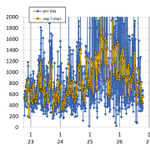
Fitbit-Aktivitätskalorien. Seit März erlebe ich eine schwierige Phase, die so wirkt, als ob die Krankheit fortschreitet. Unklar warum, unklar auch, warum 2025 zeitweise besser war.
Es quält mich so sehr, vor allem die Benommenheit und der Kopfdruck.

faei retweeted

Grateful to see the announcement of FLINTLOCK Phase 2 RCT with VYD2311 (monoclonal antibody against spike) in Long COVID and post-COVID vaccination syndrome. It will help us learn whether spike-directed therapies are helpful and for which patient subset
investors.invivyd.com/news-r…
faei retweeted

People talk about PEM like it’s just a bout of fatigue. It’s your body literally shutting down because you dared to use it. It’s microdosing death. It’s dying without the mercy of being dead.
Interessant

For MCAS and Long COVID, Japan has a Solution called Ketas (ibudilast) since 1989. The same thing is now in Western trials for neuroinflammatory disease.
Ketas is a brain-penetrant inhibitor of PDE4 (phosphodiesterase 4, the enzyme that breaks down cAMP in immune cells) and PDE10 (phosphodiesterase 10, which breaks down cAMP and cGMP in neurons), with additional activity at PDE3 and PDE11, two more enzymes that clear the same cyclic nucleotides.
It is also an allosteric inhibitor of MIF (macrophage migration inhibitory factor, a cytokine that keeps inflammation from resolving) and an antagonist at TLR4 (toll-like receptor 4, the sensor for endotoxin and damage signals).
Japan approved it for asthma and for dizziness tied to poor cerebral blood flow. The FDA has not approved it. MediciNova runs it as MN-166, with Fast Track and orphan status in ALS (amyotrophic lateral sclerosis), and it has been taken into lingering-COVID protocols for the same glial and cytokine reasons.
MCAS:
Mast cells keep cAMP (cyclic adenosine monophosphate, the signal that tells the cell to stay quiet) low through PDE4.
Low cAMP favors degranulation, TNF (tumor necrosis factor, a core inflammatory cytokine), IL-6 (interleukin-6, the fever and fatigue cytokine), and the leukotriene arm.
Ibudilast raises cAMP by blocking that enzyme, so the cell is less willing to dump. Older pharmacology also gives it leukotriene-receptor antagonism, which is the part an H1 blocker (histamine H1 receptor blocker) never covers.
MIF is released by mast cells and then calls more inflammation in. Blocking MIF shortens that loop. TLR4 is how endotoxin and mold fragments prime the mast cell before a food arrives.
Ibudilast antagonizes TLR4 and cuts the MyD88 (the adaptor that carries the TLR4 signal inward) and NF-kB (nuclear factor kappa B, the transcription switch for inflammatory genes) traffic underneath it.
Long COVID:
The persistent form is not a missing antihistamine. It is microglia that stayed on, TLR4 still ligated by residual inflammatory signals, and mast cells firing on a lowered threshold. Ibudilast crosses into the brain, suppresses glial activation, and raises cerebral blood flow.
That is the cognitive half: processing speed and the wired-but-exhausted feeling track perfusion and glial noise more than they track dopamine.
MIF and TLR4 are the systemic half. They keep the cytokine set point high after the virus is gone. PDE4 blockade in immune cells pulls TNF, IL-1β (interleukin-1 beta, the inflammasome cytokine), and IL-6 down.
People whose long COVID looks like MCAS, POTS-adjacent flares (postural orthostatic tachycardia, the heart-rate jump on standing), brain fog, and post-exertional inflammatory crashes are the overlap this molecule was built for, even though the Japanese label never used those words.


faei retweeted

Kim Linke kämpfte jahrelang gegen eine Krankheit, die manche Mediziner noch immer nicht anerkennen: ME/CFS. Dann kämpfte sie darum, sterben zu können. Sie wurde 25 Jahre alt. Ihre Eltern erzählen, was sie bis zum Ende durchmachen musste. spiegel.de/gesundheit/sterbe…

faei retweeted

Could tiny electrical gates in our nerve cells hold a clue to #LongCOVID?
➡️ A large analysis of RNA-sequencing data found 715 genes altered in LongCOVID, including 18 ion-channel genes, with several potassium channels strongly linked to immune signaling, inflammation, lipid metabolism and blood-clotting pathways. 1/

faei retweeted

Neuer, spannender Preprint von Prof Wirth @mitodicure
Orthostatic Dysfunction and Myalgic
Encephalomyelitis/Chronic Fatigue
Syndrome: A Close Reciprocal
Relationship Beyond Cardiac Preload
Failure and Hypoperfusion
preprints.org/frontend/manus…